


Abstract: Magnetic-activated cell sorting(MACS) or flow cytometry usually uses EDTA or heparin sodium-anticoagulated peripheral whole blood as the sample. Two pre-treatment methods for preparing cells: a. lyse red blood cells with hemolysin. Nuclear cells are retained for further analysis; b. density gradient centrifugation(e.g. Ficoll or Percoll) separates peripheral blood mononuclear cells(PBMCs). Centrifugation forms density stratification where the target cell layer is collected. After washing and staining, further experiment can be conducted. Choose the suitable method according to experimental requirements.
Keywords: Preparing Cells, Flow cytometry, Red blood cell lysis, Ficoll density gradient centrifugation
1. Red Blood Cell Lysis
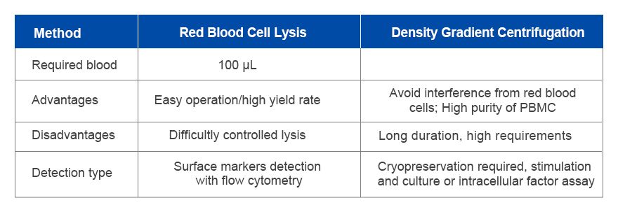
Red blood cell lysis is the specific lysis for a large number of red blood cells in the blood.(10-100 times greater than white blood cells), reducing interference with analysis for white blood cell population. The principle is that lysis buffer targeted-antigen on the surface of red blood cells can swell, deform and rupture. Effects on white blood cells are less. This method is easy-to-operate and can be completed within 10min, maintaining better integration of various white blood cells. Cell yield is also higher. However, time control of red blood cell lysis is strict. Short duration can induce incomplete lysis. Longer lysis may injure white blood cells and decrease cell viability, especially lower than viability generated by density gradient centrifugation.
1.1. Experimental Procedure
1) Add human or mouse derived peripheral blood into anticoagulative tube and store at room temperature; It's suggested to treat collected blood within 2h. Otherwise, store at 4℃ and use within 12h.
2) Take 100 μL fresh anticoagulated whole blood. Add 900 μL 1x red blood cell lysate. Mix well via gentle pipetting. Lyse for 4-15min at room temperature(human blood: 10-15 min, mouse blood: 4-5min).
3) Add 4 mL PBS. Gently mix to resuspend cells;
4) Centrifuge for 5min at 350×g at 4℃. Discard supernatant carefully.
5) Count on living cells. Adjust the required concentration with cell staining buffer and then resuspend.
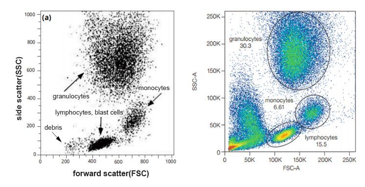
1.2. Notes
Focus on potential effects of red blood cell lysate containing fixatives on flow cytometry assay.
First, fixatives(e.g. formaldehyde or paraformaldehyde) may induce quenching of fluorescent dyes and obvious decrease of signal intensity, especially for tandem dyes(e.g. PE-Cy7, APC-Cy7 etc).
Second, fixation before antibody staining may damage or mask some antigenic epitopes due to protein crosslink or conformational change. Blocked antibody binding and recognition cause false negative or weak signal.
Thus, for weakly expressed antigen or sensitive fluorescent dyes, it's suggested to choose lysis buffer without fixatives. Try to complete surface staining before fixation.
2. Density Gradient Centrifugation
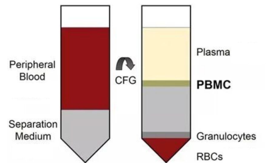
Ficoll density gradient centrifugation is the classical method for separating peripheral blood mononuclear cells(PBMCs).
Ficoll mainly consists of Ficoll 400 and meglumin diatrizoate. The average molecular weight is about 400,000. The density is usually 1.077 g/mL(human blood) or 1.084 g/mL(mouse blood). The osmotic pressure is closer to physiological conditions, unable to pass through the cell membrane.
After centrifugation, blood components are stratified according to density:
Plasma and platelets are on the top layer. Red blood cells and granulocytes sink into the tube bottom. PBMCs(density: 1.075 g/mL) are enriched at the interfaces between the plasma and Ficoll, and easy to be collected.
Notes:
Ficoll should be equilibrated to room temperature. Low temperature can induce dispersion of buffy coat and affect recovery. Besides, refer to operational specifications in the manual to ensure separation effects and cell viability.
This method is gentle and highly efficient, suitable for immunophenotype, function or culture assay.
3. Comparison among Methods for Preparing Cells
During immunophenotyping analysis with flow cytometry, whole blood sample is widely used. Easy operation can maintain the integrity of cell structure and reduce cell loss. However, proportion of red blood cells and nuclear cells in the whole blood is about 600:1, depending on sample source and species. Common sample size during detecting rare cell subpopulations may not collect sufficient events. Thus, it's suggested to increase sample volume to improve detection rate of rare cells and data reliability.
4. Applications
Red blood cell lysis is suitable for rapid flow cytometry assay of many leukocytic surface markers(especially immunophenotyping analysis without functional assay). Ficoll density gradient centrifugation is applied in PBMC experiment with high purity and activity, e.g. cell culture, functional validation, cytokine stimulation or single-cell omics analysis.
| Recommended Products | |||
| Species | Cell Populations | Flow Cytometry Antibody Combination | Cat.No |
| Human | T/B/NK cell populations detection | CD45-PerCP | PCP-30039 |
| CD3-FITC | FITC-30004 | ||
| CD16-PE | PE-30061 | ||
| CD56-PE | PE-30008 | ||
| CD19-APC | APC-30066 | ||
| Human | Thl/Th2 cell populations detection | CD3-PerCP/Cyanine5.5 | PCP55-30004 |
| CD4-FITC | FITC-30005 | ||
| IFN-γ-PE | PE-30053 | ||
| IL4-APC | APC-30043 | ||
| Mouse | Thl/Th2 cell populations detection | CD3-PerCP/Cyanine5.5 | PCP55-30002 |
| CD4-FITC | FITC-30128 | ||
| IFN-γ-PE | PE-30074 | ||
| IL4-APC | APC-30026 | ||
| Human | Treg cell populations detection | CD4-FITC | FITC-30005 |
| CD25-PE | PE-30035 | ||
| CD3-PerCP-Cy5.5 | PCP55-30004 | ||
| CD127-FineTest®647 | F647-30033 | ||
| Mouse | Treg cell populations detection | CD4-FITC | FITC-30128 |
| CD25-APC | APC-30017 | ||
| FOXP3-PE | PE-30111 | ||
REFERENCES
[1]Mass Cytometry Reveals Comparable Proportions of Leukocyte Subsets and Cell Surface P2X7 or CD39 on Human Peripheral Blood Mononuclear Cell Samples Isolated by Ficoll-Paque or SepMate Tube Density Gradient Centrifugation, PMID: 42121896.
[2]Red Blood Cell Glycation Triggers In Vivo Cerebral Erythrophagocytosis in Adult Zebrafish in a Model Mimicking Hemorrhagic Stroke, PMID: 41493138.
[3]A high-yield technique for preparing cells fixed in suspension for scanning electron microscopy, PMID: 1104641.