


Abstract: In flow cytometry, staining for intracellular protein target usually requires for fixation and permeabilization buffer kit. Fixation can stabilize cell structure and maintain morphological integrity. Permeabilization produces micropore on cell membrane, enabling entry of antibodies into cells to bind with the target protein. This process effectively exposes intracellular antigen, facilitating specific staining and detection. The fluorescent signal(e.g. surface labeled antibodies) of proteins bound on membrane surface can be reserved by using compatible fixation and permeabilization buffer kit. Proper selection of fixation and permeabilization system is very important for synchronous analysis of intracellular and superficial proteins.
Keywords: Fixation and Permeabilization, Flow Cytometry, Foxp3 Intracellular Staining, Fluorescent Signal
1. Fluorescent Signal Loss
Researches show the fluorescent signal of EYFP(CD4-Cre x Rosa26-EYFP(CD4/EYFP)) in the untreated mouse living splenocyte is the strongest. After using Foxp3 staining kit(including Fix/Perm and Perm method), fluorescence of EYFP is almost lost. Pre-fixation with 2% PFA before staining can effectively maintain EYFP signal.
This phenomenon shows fluorescence loss is induced by insufficient fixation of Fix/Perm buffer instead of fluorescent protein degradation. Soluble cytosolic EYFP is eluted from cells during permeabilization and washing steps.
2. Experimental Trials
To solve fluorescent signal loss during transcription factor(TF) staining, researchers compare different fixation/permeabilization solutions in mouse CD4-Cre x EYFP splenocyte. Further experiment shows protein content in Fix/Perm treated - supernatant is obviously higher than Triton X-100 group. Anti‑GFP capture antibody can be used to detect EYFP in Perm treated - supernatant, but can’t detect EYFP in Fix/Perm treated - sample. Leakage of EYFP is induced by insufficient fixation during Fix/Perm step. These results show fluorescent loss is induced by low fixation intensity instead of fluorophores degrade.
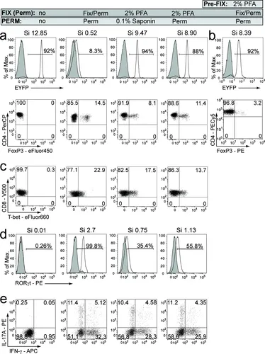
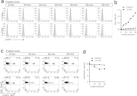
Although fixation with 2% PFA can efficiently maintain EYFP fluorescence, staining intensity of FoxP3 and proportion of positive cells obviously decreases. Staining of T-bet in CD8⁺ T cell also reduces. In double positive(DP) thymocyte consistently expressing RORγt, fixation with PFA severely reduces RORγt signal. However, in CNS-infiltrating CD4⁺ T cells from EAE mice, various intracellular cytokine staining methods for IFN‑γ and IL‑17A show similar results. In order to balance fluorescent retention and transcription factor detection, researchers decrease concentration of PFA and adjust fixation duration, then permeabilizing with Perm buffer. Longer fixation can improve EYFP signal. Fluorescence recovery is limited. Obvious FoxP3 staining loss shows it's difficult to keep the balance by regulation of PFA condition.
3. Key Breakthroughs
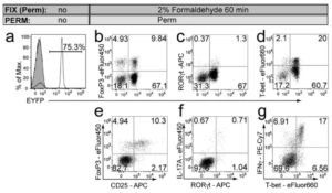
Researchers try to use a little 2% FA fixative prepared by liquid formaldehyde. After fixing for 40-60 min, the staining effect with permeabilization is better. Fluorescent signal of EYFP is clear. Positive rate is the same as untreated group. Staining efficiency of FoxP3, T-bet and RORγt completely recovers. Intensity can compare favorably with standard solution. Besides, cellular FSC/SSC morphology is better. Retention of surface markers(e.g. CD4, CD8) is more complete. The recovery is better than commercial kits. This method is also suitable for tdRFP mouse. In EAE model, 91% CNS-infiltrating CD4⁺ T cells still keep RFP positive. Expression of RORγt, IL-17A and T-bet is also detectable.
4. Complete Operation Process
Detection of Cytokines:
First, stimulate cells for 4h with PMA(50 ng/mL) and ionomycin(500 ng/mL). Add Brefeldin A(1 μg/mL) to block protein secretion.
Then surface staining: treat with Fc blocker(2.4G2, 5 μg/mL) for 10min. Incubate for 15min at 4℃ in the dark. Prepare antibodies with DPBS buffer containing 1% BSA and 0.1% NaN₃. Common markers include CD90.2, CD4, CD8, CD25, CD11b and CD45.2 etc.
Fixation: fix with 2% formaldehyde(FA, diluted by 4% Roti-Histofix with <3% formaldehyde ). Incubate for 40-60min at room temperature. Permeabilization and intracellular staining uses 0.1% saponin buffer or 1x Perm buffer. Add antibodies against FoxP3, T-bet, RORγt, IL-17A or IFN-γ etc. Staining for 30min overnight in the dark.
Finally, wash twice with the same Perm buffer. After re-suspension, collect data with the flow cytometer. This solution considers both stability of fluorescent reporter and staining effects of nuclear/cytoplasmic proteins, and is suitable for multi-parameter immunophenotyping analysis.
| Recommended Products | |||
| Species | Cell Populations | Flow Cytometry Antibody Combination | Cat.No |
| Human | T/B/NK cell populations detection | CD45-PerCP | PCP-30039 |
| CD3-FITC | FITC-30004 | ||
| CD16-PE | PE-30061 | ||
| CD56-PE | PE-30008 | ||
| CD19-APC | APC-30066 | ||
| Human | Thl/Th2 cell populations detection | CD3-PerCP/Cyanine5.5 | PCP55-30004 |
| CD4-FITC | FITC-30005 | ||
| IFN-γ-PE | PE-30053 | ||
| IL4-APC | APC-30043 | ||
| Mouse | Thl/Th2 cell populations detection | CD3-PerCP/Cyanine5.5 | PCP55-30002 |
| CD4-FITC | FITC-30128 | ||
| IFN-γ-PE | PE-30074 | ||
| IL4-APC | APC-30026 | ||
| Human | Treg cell populations detection | CD4-FITC | FITC-30005 |
| CD25-PE | PE-30035 | ||
| CD3-PerCP-Cy5.5 | PCP55-30004 | ||
| CD127-FineTest®647 | F647-30033 | ||
| Mouse | Treg cell populations detection | CD4-FITC | FITC-30128 |
| CD25-APC | APC-30017 | ||
| FOXP3-PE | PE-30111 | ||
REFERENCES
[1]A fixation-compatible protocol for intracellular and surface marker-based detection of circulating tumor cells in hepatocellular carcinoma, PMID: 41203725.
[2]Protocol to study internalization and localization dynamics of exogenously added proteins in detergent-permeabilized human cells, PMID: 40742814.
[3]Assessing the limitations of paraformaldehyde fixation for accurate cell surface receptor measurement, PMID: 41953194.