


Abstract: Apoptosis is the gene programmed active cell death, totally different from necrosis. Changes of key molecules are found during the process, e.g. externalized phosphatidylserine, decreased mitochondrial membrane potential, activated cascade of caspase and DNA fragmentation. Specific fluorescent probe for flow cytometry can accurately target and recognize molecular signal during different apoptotic phases. Quantitative apoptosis analysis of fluorescent signal can effectively distinguish among living cells, early and late apoptotic cells and necrotic cells, providing reliable evidences for objectively estimating death state of cell populations.
Keywords: Apoptosis Analysis, Flow Cytometry for Apoptosis, Apoptosis Assay, Cell Cycle Analysis
Based on principle and early and late apoptosis, the most commonly used detection methods for apoptosis analysis are specified below.
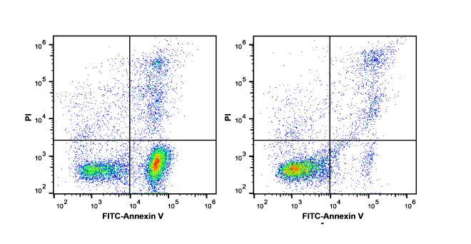
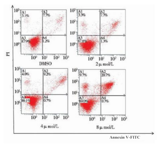
1. Annexin V and PI Staining
This is the widely used apoptosis assay method, based on externalized phosphatidylserine(PS) and changes of cell membrane integrality.
Principle:
Annexin V: a calcium-dependent phospholipid-binding protein binds to exposed PS outside cell membrane with high affinity(early apoptotic marker).
PI(Propidium Iodide): a nucleic acid dye, unable to penetrate the complete cell membrane, but accessible to damaged membrane of late apoptotic or necrotic cells.
Result interpretation(Four-quadrant analysis):
Annexin V⁻ / PI⁻: living cells
Annexin V⁺ / PI⁻: early apoptotic cells(externalized phosphatidylserine with complete membrane)
Annexin V⁺ / PI⁺: late apoptotic cells(externalized phosphatidylserine with increased membrane permeability)
Annexin V⁻ / PI⁺: necrotic cells(usually induced by mechanical damage, normal phosphatidylserine)
2. Mitochondrial Membrane Potential Detection(Early Warning)
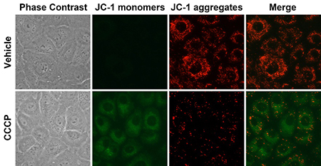
Decrease of mitochondrial membrane potential is the earliest event during apoptotic cascade, earlier than externalized phosphatidylserine and DNA breakage.
Principle: use lipophilic cationic fluorescent dyes(e.g. JC-1, TMRM, TMRE)
JC-1: It forms J-aggregates in normal mitochondria(high potential) and emits red fluorescence; its monomeric form exists in apoptotic cells(low potential) and emits green fluorescence.
Advantages: Earlier detection of apoptotic signs than Annexin V, especially suitable for studying apoptotic mechanism of mitochondrial pathway.
3. Detection of Caspase Activity(Molecular Mechanism)
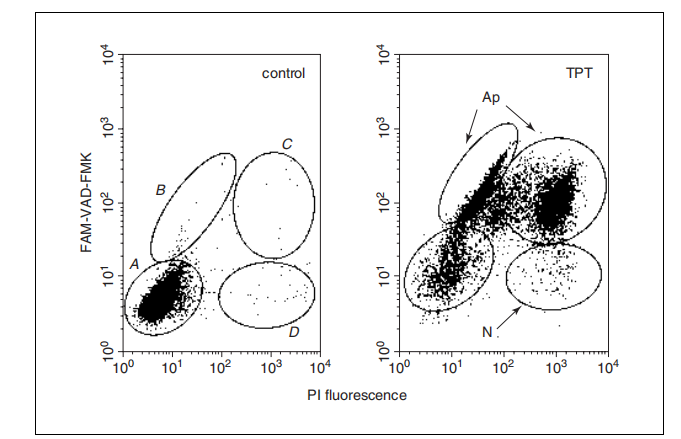
Activation of caspase family(e.g. caspase-3, -7, -9) is molecular switches for apoptotic execution.
Principle:
FLICA probe: Fluorescence-labeled caspase inhibitor covalently binds with activated caspase via entering cells. Fluorescent signal is retained in cells.
Antibody-based assay: Detect cleaved fragment of caspase with specific antibodies.(Fixation and permeabilization are required.)
Application: Deep investigation on detailed apoptotic pathways(e.g. endogenous or exogenous pathway)
4. Analysis of DNA Content(Late-stage/end-point assay)
Mainly applied in detecting DNA breakage and degradation.
Sub-G1 peak detection: DNA of apoptotic cells is lost. After ethanol fixation and PI staining, DNA content lower than G1 phase(Sub-G1) during cell cycle analysis appears.
TUNEL assay: label break ends(3'-OH) of DNA with terminal deoxynucleotidyl transferase. High specificity plays an important role in late apoptotic detection.
5. Standard Detection Method(e.g. Annexin V/PI)
During Annexin V/PI assay, sample preparation is very important. Adherent cells are gently digested with EDTA-free trypsin to prevent false positive. Suspension cells are washed via low speed centrifugation. Finally, concentration is adjusted to 1x10⁶ cells/mL. During staining, add Annexin V-FITC and PI into cell suspension respectively. Incubate at room temperature in the dark. Blank, negative, and single positive controls are required for voltage regulation, baseline determination and compensation setup during the assay. During data analysis, gating via bivariate dot plot counts cell proportion in each quadrant. Total apoptotic rate objectively reflects differences among groups.
| Recommended Products | |||
| Species | Cell Populations | Flow Cytometry Antibody Combination | Cat.No |
| Human | T/B/NK cell populations detection | CD45-PerCP | PCP-30039 |
| CD3-FITC | FITC-30004 | ||
| CD16-PE | PE-30061 | ||
| CD56-PE | PE-30008 | ||
| CD19-APC | APC-30066 | ||
| Human | Thl/Th2 cell populations detection | CD3-PerCP/Cyanine5.5 | PCP55-30004 |
| CD4-FITC | FITC-30005 | ||
| IFN-γ-PE | PE-30053 | ||
| IL4-APC | APC-30043 | ||
| Mouse | Thl/Th2 cell populations detection | CD3-PerCP/Cyanine5.5 | PCP55-30002 |
| CD4-FITC | FITC-30128 | ||
| IFN-γ-PE | PE-30074 | ||
| IL4-APC | APC-30026 | ||
| Human | Treg cell populations detection | CD4-FITC | FITC-30005 |
| CD25-PE | PE-30035 | ||
| CD3-PerCP-Cy5.5 | PCP55-30004 | ||
| CD127-FineTest®647 | F647-30033 | ||
| Mouse | Treg cell populations detection | CD4-FITC | FITC-30128 |
| CD25-APC | APC-30017 | ||
| FOXP3-PE | PE-30111 | ||
REFERENCES
[1]Refined annexin V/PI apoptosis protocol to minimize false-positive necrotic cells, PMID: 42571839.
[2]MKRN2 attenuates LPS-induced apoptosis in lung epithelial cells via ubiquitination-mediated p53 degradation, PMID: 40885043.