


Abstract: Flow cytometry is the most advanced cell quantitative analysis technology with high specificity, sensitivity and rapid detection for multi-parameter. Wide clinical applications include immunophenotyping for leukemia and lymphoma and therapeutic evaluation for hematopoietic stem cell transplantation. Roles of cellular immunophenotyping analysis are very important, including single cell suspension preparation, cell surface staining, intracellular or intranuclear antigen staining(e.g. transcription factors, cytokines). Antibody staining steps and notes will be specified below.
Keywords: Antibody Staining, Flow Cytometry, Cell Quantitative Analysis, Immunophenotyping
1. Flow Cytometry Analysis for Immune Cells
Wide scientific and clinical applications mainly cover the following project:
1.1. Basic Analysis for Lymphocyte Subpopulation
Evaluate immune homeostasis, monitor HIV/AIDS and screen immunodeficiency, including T cell(CD3⁺CD4⁺/CD8⁺), B cell(CD19⁺/CD20⁺) and NK cell(CD3⁻CD56⁺/CD16⁺).
1.2. Functional Phenotyping of T/B Cell Subpopulation
Helpful to analysis for autoimmune diseases, infection or vaccine response mechanism, e.g. Th1/Th2/Th17/Treg(identify via transcription factors or cytokines, like T-bet/GATA3/RORγt/Foxp3 etc), Memory B cell(IgD/CD27) and plasmablast(CD38ʰⁱCD27ʰⁱ).
1.3. Activation, Depletion and Evaluation for Proliferative State
Detection of activated markers(CD25, HLA-DR), immune checkpoints(PD-1, TIM-3, LAG-3) and proliferative markers(Ki-67, CFSE) plays an important role in tumor immunotherapy.
1.4. Analysis for Cytokines and Apoptosis
Detect secretion profile of IFN-γ, IL-17 via intracellular staining. Alternatively evaluate apoptosis with Annexin V/PI.
1.5. Clinical Applications
Multicolor flow cytometry can accurately perform high-dimensional analysis for single-cell level, strongly supporting pathogenetic mechanism, diagnostic typing and therapeutic evaluation, e.g. immunophenotyping for leukemia/lymphoma, restriction analysis for κ/λ light chain(determine clonality of B cell), CAR-T cell monitoring and diagnosis of primary immunodeficiency.
2. Antibody Staining Steps
2.1. Surface Antibody Staining
Sample preparation during flow cytometry includes cell suspension and whole blood. Adjust concentration of cell suspension to 1×10⁷ cells/mL. Add 100 μL into surface antibodies and incubate for 30min at 2-8℃ in the dark; Then, add 1-2 mL staining buffer. Centrifuge for 5min at 300×g and discard supernatant. Resuspend with 200 μL buffer for further detection. Add antibodies into anticoagulated whole blood(human: 100 μL, mouse: 30-50 μL) following the same incubation step. Add red blood cell lysate to remove red blood cells. After centrifugation and washing, resuspend with 200 μl buffer again.
2.2. Intracellular Staining of Antibodies
Take Intracellular Fixation/Permeabilization Buffer Kit (Ready To Use) -- Cat.No: K082R for example:
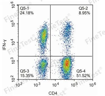
During flow cytometry assay for intracellular antigen, add about 1×10⁶(100 μL) into FACS tube. Viability staining and blocking for Fc receptor are optional. Then, add fluorescence-labeled antibodies to stain cell surface markers. After incubation, resuspend with 1 mL PBS containing 1% BSA. Centrifuge for 5min at 300×g and discard supernatant. Then, resuspend with 200 µL PBS containing 1% BSA. Add equal volume of fixation buffer and fix for 30-60min at room temperature in the dark.(60min is suggested for 25℃ below.) Centrifuge at 600×g and discard supernatant. Wash with 1 mL PBS containing 1% BSA. Store cells at 4℃ overnight. In the next day, centrifuge and add 100µL permeabilization buffer to resuspend cells. Add intracellular staining antibodies and incubate for 30min at room temperature in the dark.
2.3. Intranuclear Antibody Staining
Take Foxp3/Transcription Factor Staining Kit -- Cat.No: K081 for example:
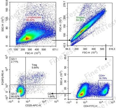
This kit can synchronously detect cytoplasm and intranuclear proteins. Add about 1×10⁶(100 μL) cells into FACS tube. Viability staining is optional for distinguishing dead or live cells. Blocking of Fc receptor depends on requirements. Then, add fluorescence-labeled surface antibodies and incubate in the dark. After incubation, add 1 mL cell staining buffer[K079]. Centrifuge for 5min at 300×g and discard supernatant. Resuspend with 100 μL buffer. Then, add 1 mL 1×Fixation Working Solution. Mix well with the vortex mixer. Fix for 30min at 4℃. Centrifuge for 5min at 600×g and discard supernatant. Wash twice with 2 mL 1×Permeabilization Working Buffer(following the centrifugation before). Finally, resuspend cells with 100 μL buffer. Add fluorescence-labeled intranuclear antibodies and incubate for 30min at room temperature in the dark. After incubation, wash with permeabilization buffer. Then, resuspend with proper amount of buffer(K079).
3. Flow Cytometry Services
FineTest is committed to provide professional flow cytometry color matching services and high-quality reagents. Scientific optimization of multicolour antibody combination effectively reduces spectral overlap and background interference, greatly improving specificity and sensitivity of detection. Accurate recognition and sorting the target immunocyte population helps researchers obtain efficient, stable, and reliable data, strongly supporting various research fields like immunology, tumor immunity, and autoimmune diseases etc.
| Recommended Products | |||
| Species | Cell Populations | Flow Cytometry Antibody Combination | Cat.No |
| Human | T/B/NK cell populations detection | CD45-PerCP | PCP-30039 |
| CD3-FITC | FITC-30004 | ||
| CD16-PE | PE-30061 | ||
| CD56-PE | PE-30008 | ||
| CD19-APC | APC-30066 | ||
| Human | Thl/Th2 cell populations detection | CD3-PerCP/Cyanine5.5 | PCP55-30004 |
| CD4-FITC | FITC-30005 | ||
| IFN-γ-PE | PE-30053 | ||
| IL4-APC | APC-30043 | ||
| Mouse | Thl/Th2 cell populations detection | CD3-PerCP/Cyanine5.5 | PCP55-30002 |
| CD4-FITC | FITC-30128 | ||
| IFN-γ-PE | PE-30074 | ||
| IL4-APC | APC-30026 | ||
| Human | Treg cell populations detection | CD4-FITC | FITC-30005 |
| CD25-PE | PE-30035 | ||
| CD3-PerCP-Cy5.5 | PCP55-30004 | ||
| CD127-FineTest®647 | F647-30033 | ||
| Mouse | Treg cell populations detection | CD4-FITC | FITC-30128 |
| CD25-APC | APC-30017 | ||
| FOXP3-PE | PE-30111 | ||
REFERENCES
[1]Flow Cytometry and Single-Cell Analysis for Characterizing Microglia Activation in Early Postnatal Mouse Brain Development, PMID: 41115126.
[2]Optimization of a 40-Color Spectral Flow Cytometry Panel for Human Blood Immunophenotyping: Impact of Blood Volume, Sample Preservation, and Overnight Staining, PMID: 41454662.