PGK1 antibody

11th Anniversary Sale
Synonyms:Phosphoglycerate kinase 1|Cell migration-inducing gene 10 protein|Primer recognition protein 2 (PRP 2)|PGK1|PGKA antibody
Catalogue No.:FNab06354Reactivity:Human, Mouse, Rat
Host:RabbitTested Application:ELISA, WB, IHC, IF
Clonality:polyclonalIsotype:IgG
Size Price
100µg Inquiry
Dispatch Time: About 3 working days
  • SPECIFICATIONS
  • CITATIONS
  • FIGURES
  • CONDITIONS
  • FAQS
Product Name
PGK1 antibody
Catalogue No.
FNab06354
Size
100μg
Form
liquid
Purification
Immunogen affinity purified
Purity
≥95% as determined by SDS-PAGE
Clonality
polyclonal
Isotype
IgG
Storage
PBS with 0.02% sodium azide and 50% glycerol pH 7.3, -20℃ for 12 months (Avoid repeated freeze / thaw cycles.)
Immunogen
Immunogen
phosphoglycerate kinase 1
Alternative Names
Phosphoglycerate kinase 1|Cell migration-inducing gene 10 protein|Primer recognition protein 2 (PRP 2)|PGK1|PGKA antibody
UniProt ID
P00558
Observed MW
47 kDa
Application
Tested Applications
ELISA, WB, IHC, IF
Recommended dilution
WB: 1:500 - 1:2000; IHC: 1:50 - 1:200; IF: 1:50 - 1:200
Validated Images
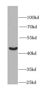
HeLa cells were subjected to SDS PAGE followed by western blot with FNab06354(PGK1 antibody) at dilution of 1:1200
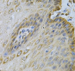
Immunohistochemistry of paraffin-embedded human esophagus using FNab06354( PGK1 Antibody) at dilution of 1:50
Background
The protein encoded by this gene is a glycolytic enzyme that catalyzes the conversion of 1, 3-diphosphoglycerate to 3-phosphoglycerate. The encoded protein may also act as a cofactor for polymerase alpha. Additionally, this protein is secreted by tumor cells where it participates in angiogenesis by functioning to reduce disulfide bonds in the serine protease, plasmin, which consequently leads to the release of the tumor blood vessel inhibitor angiostatin. The encoded protein has been identified as a moonlighting protein based on its ability to perform mechanistically distinct functions. Deficiency of the enzyme is associated with a wide range of clinical phenotypes hemolytic anemia and neurological impairment. Pseudogenes of this gene have been defined on chromosomes 19, 21 and the X chromosome.
IF: 17.388
Journal:
Journal of Hematology & Oncology
Cited Date:
2022-06-10
Product:
How many times can antibodies be recycled?

First, usually it's not suggested to recycle antibodies. After use, buffer system of antibodies has changed. The storage condition of recycled antibodies for different customers also varies. Thus, the performance efficiency of recycled antibodies can’t be guaranteed. Besides, FineTest ever conducted the antibody recycling assay. Assay results show recycling times of different antibodies also varies. Usually, higher antibody titer allows more repeated use. Customers can determine based on experimental requirements.

Notes: After incubation, we recycle rest antibodies to centrifuge tube and store at 4℃. High titer antibodies can be stored for a minimum of one week. Reuse about three times.

What are components of FineTest antibody buffer?

Components of FineTest antibody buffer are usually PBS with proclin300 or sodium azide, BSA, 50% glycerol. Common preservative is proclin300 or sodium azide, which is widely applied in the lab and industry.

How about the storage temperature and duration of FineTest antibodies?

Most antibodies are stored at -20℃. Directly-labeled flow cytometry antibodies should be stored at 2 - 8℃. The shelf life is one year. If after sales issues for purchased antibodies appear, return or replacement is available. Usually, antibodies can be still used after the one-year warranty. We can offer technical support services.

Is dilution required for FineTest antibodies? What’s the dilute solution?

Directly-labeled flow cytometry antibodies are ready-to-use without dilution. Other antibodies are usually concentrated. Follow the dilution ratio suggested in the manual. Dilute solution for different experiments also varies. Common antibody dilution buffers are acceptable(e.g. PBST, TBST, antibody blocking buffer).

How to retrieve antibodies for immunohistochemistry?

Common retrieval buffers: Tris-EDTA Buffer(pH 9.0); Citrate Buffer(pH 6.0)

Heat induced antibody retrieval:

Method 1: Water-bath heating: Put the beaker with retrieval buffer and slide in the boiling water bath. Keep the boiling state for 15min. Naturally cool to room temperature;

Method 2: Microwave retrieval: Put the beaker with retrieval buffer and slide in the microwave oven. Heat at high power for 5min, Switch OFF for 3min, Heat at medium power for 5min. Naturally cool to room temperature.

How to choose secondary antibodies?

(1) Secondary antibodies react with primary antibodies. Thus, secondary antibodies should be against host species of primary antibodies. E.g. If the primary antibody is derived from rabbit, the relevant secondary antibody should be against rabbit. E.g. goat anti rabbit or donkey anti rabbit.

(2) Choose secondary antibody conjugates according to the experimental type, e.g. ELISA, WB, IHC etc. Common enzyme conjugated secondary antibodies are labelled by HRP, AP etc. Fluorescin or dye labelled secondary antibodies are applied in immunofluorescence and flow cytometry(e.g. FITC, Cy3).

Product Comparison