BAX antibody

Synonyms:Apoptosis regulator BAX|Bcl-2-like protein 4 (Bcl2-L-4)|BAX|BCL2L4 antibody
Catalogue No.:FNab00810Reactivity:Human, Mouse, Rat, Rabbit, Sheep
Host:RabbitTested Application:ELISA, WB, IHC, IP, FC
Clonality:polyclonalIsotype:IgG
Size Price
100µg Inquiry
Dispatch Time: About 3 working days
  • SPECIFICATIONS
  • CITATIONS
  • FIGURES
  • CONDITIONS
  • FAQS
Product Name
BAX antibody
Catalogue No.
FNab00810
Size
100μg
Form
liquid
Purification
Immunogen affinity purified
Purity
≥95% as determined by SDS-PAGE
Clonality
polyclonal
Isotype
IgG
Storage
PBS with 0.02% sodium azide and 50% glycerol pH 7.3, -20℃ for 12 months(Avoid repeated freeze / thaw cycles.)
Immunogen
Immunogen
BCL2-associated X protein
Alternative Names
Apoptosis regulator BAX|Bcl-2-like protein 4 (Bcl2-L-4)|BAX|BCL2L4 antibody
UniProt ID
Q07812
Observed MW
21 kDa
Application
Tested Applications
ELISA, WB, IHC, IP, FC
Recommended dilution
WB: 1:1000-1:10000; IP: 1:1000-1:10000; IHC: 1:20-1:200
Validated Images
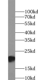
HEK-293 cells were subjected to SDS PAGE followed by western blot with FNab00810(BAX antibody) at dilution of 1:1000
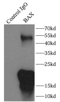
IP Result of anti-BAX (IP: FNab00810, 3ug; Detection: FNab00810 1:2000) with Raji cells lysate 3500ug.
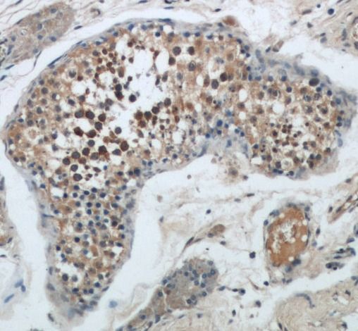
Immunohistochemistry of paraffin-embedded human kidney tissue slide using FNab00810(BAX Antibody) at dilution of 1:50
Background
BAX, also named as BCL2L4, is a pro-apoptotic member of the Bcl-2 protein family, which plays a pivotal role in controlling cell life and death. Bax largely localizes to the cytoplasm of healthy cells, but accumulates on the outer mitochondrial membrane upon apoptosis induction(PMID: 9108035). BAX can commit a cell to apoptosis by translocation from the cytosol to the mitochondria and permeabilization of the outer mitochondrial membrane, which leads to the release of cytochrome c from mitochondria(PMID: 21763611). The expression of BAX is upregulated by the tumor suppressor protein p53, and BAX has been shown to be involved in p53-mediated apoptosis(PMID: 8183579).
IF: 3.4
Journal:
Molecular Medicine Reports
Author:
Spinal Surgery Department 2, Hebei Medical University Third Hospital, Shijiazhuang, Hebei 050011, P.R. China.
Cited Date:
2025-03-14
Product:
IF: 2.6
Journal:
Drug and Chemical Toxicology
Author:
Department of Physiology, Faculty of Medicine, Firat University, Elazig, Turkey
Cited Date:
2023-09-15
Product:
IF: 0.8
Journal:
Anatomia, Histologia, Embryologia
Author:
Department of Histology and Embryology, Faculty of Medicine, Hitit University, Corum, Turkey.
Cited Date:
2024-08-23
Product:
How many times can antibodies be recycled?

First, usually it's not suggested to recycle antibodies. After use, buffer system of antibodies has changed. The storage condition of recycled antibodies for different customers also varies. Thus, the performance efficiency of recycled antibodies can’t be guaranteed. Besides, FineTest ever conducted the antibody recycling assay. Assay results show recycling times of different antibodies also varies. Usually, higher antibody titer allows more repeated use. Customers can determine based on experimental requirements.

Notes: After incubation, we recycle rest antibodies to centrifuge tube and store at 4℃. High titer antibodies can be stored for a minimum of one week. Reuse about three times.

What are components of FineTest antibody buffer?

Components of FineTest antibody buffer are usually PBS with proclin300 or sodium azide, BSA, 50% glycerol. Common preservative is proclin300 or sodium azide, which is widely applied in the lab and industry.

How about the storage temperature and duration of FineTest antibodies?

Most antibodies are stored at -20℃. Directly-labeled flow cytometry antibodies should be stored at 2 - 8℃. The shelf life is one year. If after sales issues for purchased antibodies appear, return or replacement is available. Usually, antibodies can be still used after the one-year warranty. We can offer technical support services.

Is dilution required for FineTest antibodies? What’s the dilute solution?

Directly-labeled flow cytometry antibodies are ready-to-use without dilution. Other antibodies are usually concentrated. Follow the dilution ratio suggested in the manual. Dilute solution for different experiments also varies. Common antibody dilution buffers are acceptable(e.g. PBST, TBST, antibody blocking buffer).

How to retrieve antibodies for immunohistochemistry?

Common retrieval buffers: Tris-EDTA Buffer(pH 9.0); Citrate Buffer(pH 6.0)

Heat induced antibody retrieval:

Method 1: Water-bath heating: Put the beaker with retrieval buffer and slide in the boiling water bath. Keep the boiling state for 15min. Naturally cool to room temperature;

Method 2: Microwave retrieval: Put the beaker with retrieval buffer and slide in the microwave oven. Heat at high power for 5min, Switch OFF for 3min, Heat at medium power for 5min. Naturally cool to room temperature.

How to choose secondary antibodies?

(1) Secondary antibodies react with primary antibodies. Thus, secondary antibodies should be against host species of primary antibodies. E.g. If the primary antibody is derived from rabbit, the relevant secondary antibody should be against rabbit. E.g. goat anti rabbit or donkey anti rabbit.

(2) Choose secondary antibody conjugates according to the experimental type, e.g. ELISA, WB, IHC etc. Common enzyme conjugated secondary antibodies are labelled by HRP, AP etc. Fluorescin or dye labelled secondary antibodies are applied in immunofluorescence and flow cytometry(e.g. FITC, Cy3).

Product Comparison