Abstract: Fluorescent proteins(FPs) are derived from marine organisms(e.g. jellyfish, coral etc). They can realize autofluorescence without exogenous substrates. Their roles in modern life science research are very important. Wide applications of fluorescent proteins in flow cytometry include labeling, gene expression tracking, protein localization and cellular dynamics. This article presents fluorescent proteins from basic features, type and applications in flow cytometry.
Keywords: Fluorescent Proteins, Flow Cytometry, Luminescent Proteins, Bioluminescence
1. Types of Fluorescent Proteins
Based on emission wavelength, six kinds of FPs include BFP, CFP, GFP, YFP, RFP and IRFP. The nomenclature has specific meaning. “Turbo” refers to rapid maturation. "e" or "E"(enhanced) refers to optimized or enhanced version. "m", "d", "t" refers to monomer, dimer and tetramer respectively. Many names are derived from organisms. E.g. DsRed is derived from Discosoma striata. Understanding of nomenclature helps to choose suitable fluorescent proteins.
2. Significance of Fluorescent Proteins in Gene Fusion
Fluorescent proteins are mainly applied in gene fusion. Co-expression of fluorescent protein gene linked with target gene can form functional and fluorescent fusion protein. Then, scientists can observe expression, localization and dynamic changes of target proteins in living cells, further evaluating transfection efficiency and gene activity. Construction of fluorescent biosensor can detect intracellular physiological parameters(e.g. pH, calcium ion, redox status etc).
3. Applications of Fluorescent Proteins in Flow Cytometry
Although performance of FPs in microimaging is high, difficulties of FPs in flow cytometry are still a lot. Residence time of cells through laser point is very short. Thus, strong fluorescent signal and high brightness are required. Specific wavelength(e.g. 488nm) is set to match with the limit excitation efficiency. The wide emission spectrum easily causes spectral overlap between different fluorescent proteins and traditional dyes. Compensation is more difficult. Thus, not all imageable fluorescent proteins are suitable for flow cytometry. Next, selection of FPs for flow cytometry assay is specified below.
3.1. GFPs
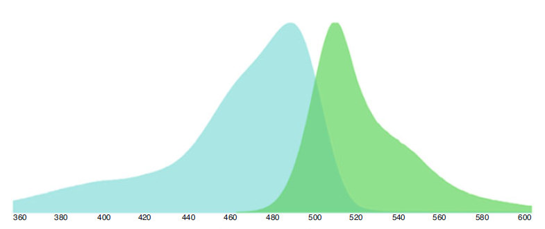
EGFP(ex488/em510) is the most commonly used FP in flow cytometry, and well-matched with 488nm laser. Performance of mEmerald and mNeonGreen is high. It's suggested to match with narrow band filters(e.g. BP510/20) to improve signal-to-noise ratio.
3.2. BFPs & CFPs
BFPs(e.g. eBFP2) requires for UV/violet excitation around 354-424 nm; CFPs(e.g. Cerulean, eCFP) can be efficiently excited by 405nm laser. AmCyan(ex458/em489) has been applied in antibody conjugation, and extended application of violet laser.
3.3. YFPs
The best excitation of Venus and Citrine is around 514–532nm, also detectable on 488nm device. Optical filters like BP550/30 are suggested for YFPs instead of GFPs.
3.4. OFPs & RFPs
Derived from corals, OFPs & RFPs(e.g. tdTomato, mCherry, mRuby etc) are suitable for excitation of 561nm laser. Tissue penetration is better. Avoid to detect mCherry with 488nm.
3.5. iRFPs
Derived from bacterial phytochrome, emission of iRFPs depends on endogenous biliverdin. Excitation requires for 630nm or above. iRFPs also provide new choices for high dimensional flow cytometry analysis.
4. Conclusion
Fluorescent proteins are key research tools in life science. Deeper understanding of their spectral properties, excitation requirements and applications is very important for multi-color panel design, device selection and data analysis. Brighter and more stable fluorescent proteins with better spectrum are expected to appear, strongly supporting high dimensional flow cytometry analysis and biomedical research.
| Recommended Products | |||
| Species | Cell Populations | Flow Cytometry Antibody Combination | Cat.No |
| Human | T/B/NK cell populations detection | CD45-PerCP | PCP-30039 |
| CD3-FITC | FITC-30004 | ||
| CD16-PE | PE-30061 | ||
| CD56-PE | PE-30008 | ||
| CD19-APC | APC-30066 | ||
| Human | Thl/Th2 cell populations detection | CD3-PerCP/Cyanine5.5 | PCP55-30004 |
| CD4-FITC | FITC-30005 | ||
| IFN-γ-PE | PE-30053 | ||
| IL4-APC | APC-30043 | ||
| Mouse | Thl/Th2 cell populations detection | CD3-PerCP/Cyanine5.5 | PCP55-30002 |
| CD4-FITC | FITC-30128 | ||
| IFN-γ-PE | PE-30074 | ||
| IL4-APC | APC-30026 | ||
| Human | Treg cell populations detection | CD4-FITC | FITC-30005 |
| CD25-PE | PE-30035 | ||
| CD3-PerCP-Cy5.5 | PCP55-30004 | ||
| CD127-FineTest®647 | F647-30033 | ||
| Mouse | Treg cell populations detection | CD4-FITC | FITC-30128 |
| CD25-APC | APC-30017 | ||
| FOXP3-PE | PE-30111 | ||
REFERENCES
[1]Novel Near-Infrared Fluorescent Protein for Biliverdin Biosensing with a Hydrogel Scaffold, PMID: 41247245.
[2]Physical Mechanisms of an Unconventional Green Fluorescent Protein Indicator for Chloride, PMID: 41784459.