


Abstract: Innate lymphoid cells(ILCs) include subsets like ILC1, ILC2, ILC3, sharing phenotype and function similar with T cells. Roles in lymphatic tissue development, tissue repair, and anti-infective immunity are important. ILCs can rapidly respond to microenvironment signal and regulate homeostasis of inflammation and immunity. The subset ratio or dysfunction is closely related to various diseases. Accurate identification of ILCs via decreasing T cell contamination is specified below.
Keywords: Innate lymphoid cells(ILCs), ILC identification, T cell contamination, Tissue-specific markers, Flow cytometry analysis
1. Developmental Pathway and Functional Plasticity of ILC1 in Immune Responses
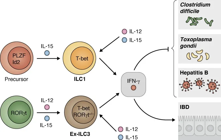
The figure below shows developmental pathway of ILC1 and its key roles in immune response. ILC1 directly differentiates from IL-15 induced PLZF⁺Id2⁺ precursors, which is driven by T-bet. Transdifferentiation of ILC3 into Ex-ILC3 is induced by IL-12 and IL-15. (expressing T-bet and low level RORγt). IFN-γ secreted by them is involved in removal of pathogens (e.g. clostridioides difficile, toxoplasma gondii and hepatitis B virus etc). Dual roles in IBD include mediation of protective immunity and aggravation of intestinal inflammation. This figure highlights plasticity of ILC1 and its key roles in infection and autoimmune regulation.
2. Effects of T Cell Contamination on ILC Identification and Experimental Reproducibility
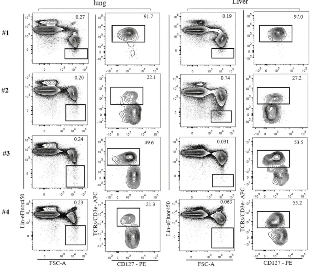
Subsets of ILC family(ILC1, ILC2, ILC3) are lineage-negative(Lin⁻) instead of expressing RAG dependent antigen receptor and identification by TCR or BCR. Lin cocktails of common mice include Ter119, Gr-1, CD11c, CD11b, CD19 and T cell markers(e.g. CD3ε/TCR), and are used to remove mature hematopoietic cells. Some cells lowly express Lin markers. If T cell markers are included in Lin cocktail, complete removal of TCR⁺ during gating is difficult. T cell contamination in ILC group is the key reason for different assay results. Residual of TCR⁺ cell is still obviously seen in Lin⁻ gating.
3. Decrease of T Cell Contamination for Improving ILC Identification
Identification of ILC is improved by decreasing T cell contamination: Remove CD3ε/TCR antibody from Lin cocktail. Labeling in independent fluorescent channel accurately eliminates TCR⁺ cells to obviously improve analysis of ILC. The assay analyzes liver, lung, spleen, and small intestine tissues via C57BL/6J, Rorγt-eGFP reporter and Rag1⁻/⁻ mouse. All tissues are specifically treated: Liver enriches immunocytes via Percoll gradient centrifugation; Lung is purified by Percoll following digestion with Collagenase IV and DNase I; Epithelial cell-free small intestine is digested by Collagenase VIII and DNase I. Then, sequential filtration obtains single cell suspension.
4. Multi-parameter Phenotyping of ILC Subsets in Murine Tissues
Accurate identification of ILC requires for tissue-specific markers and transcription factors. In B6 mouse liver, ILC1 is defined as Lin⁻ NKp46⁺ NK1.1⁺ TCR⁻ CD49a⁺ CD49b⁻(Eomes⁻T-bet⁺RORγt⁻). Avoid cross-expression of CD49a in T/NKT cells and dependence on Thy1 or CD127. Phenotype of ILC2 varies among tissues: ILC2 in lung is usually ST2⁺. ILC2 in small intestine is usually ST2⁻, which highly express KLRG1 and IL-17RB; ILC2 in liver is composed of ST2⁺/⁻. Identification of ILC3 depends on RORγt(or Rorγt-eGFP reporter) since the high overlap of surface markers. Typical phenotypes are Lin⁻ Thy1⁺ CD127⁺ TCR⁻ RORγt⁺, some of which express NKp46. NK1.1 negative can be distinguished from ILC1/NK. ILC3 in wild mouse lung is lower, but obviously increases in Rag1⁻/⁻ mouse. Multi-color flow cytometry and staining of transcription factor play an important role in accurate analysis of ILC subsets.
5. Strategies for Accurate ILC Identification
Accurate identification of ILC should strictly remove T cell contamination. It's suggested to stain CD3ε/TCR and Lin cocktail in separated channels. Different expression of ILC markers in tissues requires for tissue-specific strategy. Roles of transcription factors(E.g. GATA3, RORγt, T-bet/Eomes) in subset identification are important; Markers like CD49a, Thy1, CD127, ST2 are not completely specific, and integrated assessment is required. Spectral flow cytometry for limited channels can improve multi-parameter ability.
| Recommended Products | |||
| Species | Cell Populations | Flow Cytometry Antibody Combination | Cat.No |
| Human | T/B/NK cell populations detection | CD45-PerCP | PCP-30039 |
| CD3-FITC | FITC-30004 | ||
| CD16-PE | PE-30061 | ||
| CD56-PE | PE-30008 | ||
| CD19-APC | APC-30066 | ||
| Human | Thl/Th2 cell populations detection | CD3-PerCP/Cyanine5.5 | PCP55-30004 |
| CD4-FITC | FITC-30005 | ||
| IFN-γ-PE | PE-30053 | ||
| IL4-APC | APC-30043 | ||
| Mouse | Thl/Th2 cell populations detection | CD3-PerCP/Cyanine5.5 | PCP55-30002 |
| CD4-FITC | FITC-30128 | ||
| IFN-γ-PE | PE-30074 | ||
| IL4-APC | APC-30026 | ||
| Human | Treg cell populations detection | CD4-FITC | FITC-30005 |
| CD25-PE | PE-30035 | ||
| CD3-PerCP-Cy5.5 | PCP55-30004 | ||
| CD127-FineTest®647 | F647-30033 | ||
| Mouse | Treg cell populations detection | CD4-FITC | FITC-30128 |
| CD25-APC | APC-30017 | ||
| FOXP3-PE | PE-30111 | ||
REFERENCES
[1]Identification, characterization, and expansion of innate lymphoid cells derived from canine peripheral blood mononuclear cells, PMID: 42528714.
[2]Development and computational analysis of high dimensional spectral flow cytometry data for the resolution of innate lymphoid cells in the mammary tumor microenvironment, PMID: 41676157.