Abstract: Copper is involved in important biological processes like cellular respiration, antioxidant response and neurotransmitter synthesis. Balance of copper is very important for cells and organ health. But excessive copper ions may cause toxic reaction, resulting in cell damage and death. Copper ions induce oxidative stress and mitochondrial dysfunction by damaging acylated tricarboxylic acid cycle protein. Cuproptosis is a special copper-induced cell death mechanism, different from traditional apoptosis, necrosis, and autophagy.
Keywords: Copper Metabolism, Copper Induced Cell Death, Cuproptosis-induced Diseases, Cuproptosis Detection
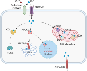
1. Copper Metabolism Pathway
In order to maintain copper homeostasis, cells exactly regulate intake, distribution, and excretion of copper by synergism of copper enzymes, copper chaperones and membrane transport protein. Normal copper level can avoid excessive and insufficient damage.
Copper metabolism pathway is briefly outlined below: Extracellular Cu2+ is first reduced to Cu+ by reductase STEAP. Cu+ enters cells by copper transporter CTR1. Cu+ is transferred to cytoplasmic copper chaperones (e.g. copper chaperone for superoxide dismutase and SOD1), and further transferred to specific subcellular compartments(e.g. mitochondrion, trans Golgi network and nucleus). In mitochondrion, copper is involved in respiratory chain and redox pathway by binding with cytochrome c oxidase(CCO). In mitochondrial intermembrane space, COX17 transfers copper to SCO1 or COX11. Then, copper is transferred to cytochrome oxidase subunit. In nucleus, copper can bind with transcription factor to drive gene expression. Finally, in TGN, Copper-ATPase transporter proteins ATP7A and ATP7B transfer copper to TGN cavity from cytoplasm, activating copper dependent enzyme in secretory pathway. When copper level in cytoplasm greatly increases, ATP7A and ATP7B leaves from TGN to promote excretion of copper.
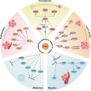
2. Signaling Pathway in Cancer
Copper is closely related to various signaling pathways by binding with and activating key molecules in these pathways. Copper plays an important role in RTK-related signaling pathway, and bind with phosphorylated RTK without dependency on ligands to result in the activation of RTK. Activated RTK then causes phosphorylation of ERK and ATK and finally promotes cell migration and proliferation. Besides, copper ion also affects PI3K-AKT pathway by directly activating PI3K, and indirectly activating AKT via binding with specific site of PDK1. The copper-induced AKT activation further catalyzes phosphorylation and redistribution of FoxO1a and FoxO4, promoting cell proliferation and growth of tumor. Meanwhile, the activation of MAPK pathway also requires copper ion. Copper promotes phosphorylation of ERK1/2 by binding with MEK1, and also activates JNK to affect development of tumor.
3. Cuproptosis-induced Diseases
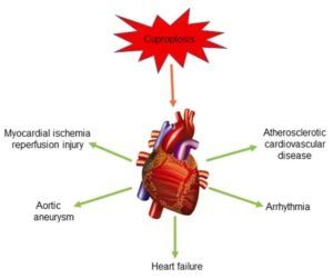
3.1. Cardiovascular Diseases
Abnormal copper metabolism is one of the important inducements for cardiovascular diseases like atherosclerosis. Copper ion is involved in oxidative modification of low density lipoprotein and promotes formation of atherosclerosis. Besides, copper ion can accelerate the development of cardiovascular diseases by inducing cuproptosis of endothelial cells and destroying the integrity of the vascular wall.
3.2. Cancer
Excessive accumulation of copper in cancer cells can mediate death of cancer cells. Thus, targeted copper therapy is very potential in cancer treatment. Cuproptosis concept depends on research on anti-tumor copper ion carriers. These carriers can transfer copper ion to cancer cells and mitochondria, inducing death of cancer cells. Besides, copper ion affects tumor development, growth, migration, angiogenesis, and metabolism by binding with and activating key molecules of various signaling pathway.
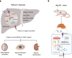
3.3. Copper Overload Diseases
Wilson's Disease is a genetic disease. Copper can’t be normally excreted from bile, resulting in excessive accumulation of copper in the body, liver injury and neurological disorder. Long-term and excessive intake of copper may also cause copper poisoning. Symptoms are nausea, vomiting, diarrhea and abdominal pain etc. In severe cases, life may be threatened.
4. Cuproptosis Detection
Cuproptosis detection methods include cell assay and molecular mechanism. Cell assays include CCK8/MTT and Annexin V/PI etc to evaluate cell viability. Measure copper content by atomic absorption spectroscopy or inductively coupled plasma mass spectrometry and observe change of cell morphology with electron microscope.
Molecular Mechanism: Detect protein level with Western blot, ELISA and IF. Measurement includes enzymatic activity of oxidative stress and TCA cycle pathways. Also analyse changes of metabolite. Using these methods together with changes in protein levels completely validate effects and mechanism of cuproptosis.
5. Recommended Products
|
Target |
Antibody |
ELISA Kit |
Recombinant Protein |
|
DLD |
|||
|
DLAT |
|||
|
LIAS |
|||
|
PDHA1 |
|||
|
PDHB |
PDHB recombinant protein |
||
|
GLS |
|||
|
GAC |
GAC recombinant protein |
||
|
CDKN2A |
|||
|
GCSH |
GCSH ELISA Kit |
||
|
FASN |
FASN recombinant protein |
||
|
DHFR |
|||
|
SLC31A1 |
|||
|
FDX1 |
REFERENCES
[1] Cuproptosis: Unraveling the Mechanisms of Copper-Induced Cell Death and Its Implication in Cancer Therapy, PMID: 38339398.
[2] Cuproptosis mediates copper-induced testicular spermatogenic cell death, PMID: 38284772.