


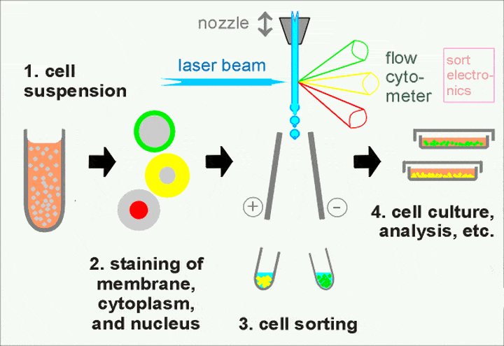
Abstract: Cell sorting is an advanced cell analysis method utilizing flow cytometry sorter. This technology detects intensity of scattered light and emitted fluorescence following excitation of high-energy laser to fluorescence labeled single cell or particle. These optical signals enable rapid and accurate qualitative and quantitative analysis for cell morphology, physiological and biochemical characteristics, immunophenotype, genetic information and molecular function. Thereby, efficient sorting for specific cell populations is widely used for life science research and clinical diagnosis.
Keywords: Cell sorting, Flow cytometry, Sample compatibility, Quality control, Cell viability
1. Applications and Advantages of Cell Sorting
Cell sorting is suitable for difficult cell separations, based on multi-color fluorescence labeling; different signal intensity and weak signal; functional status(e.g. endocytosis, apoptosis etc) or intracellular markers(e.g. DNA content). This technology can obtain high purity target cell(95%-100%), facilitating precise dispensing of single-cell into plates. Besides, rare cells can be enriched from low abundance sample(e.g. 0.001%). Sorting potential new subsets with unknown features is the powerful tool exploring cell heterogeneity.
2. High Sample Compatibility of Cell Sorting
High compatibility for samples shows applicability of cell sorting for various biomaterials. Main sample types include: liquid bone marrow and peripheral blood for single cell suspension preparation; Solid tissue block can be tested after digestion; In vitro culture of cell lines is the common experimental model; Various exfoliated cells(e.g. pleural and ascitic fluid, brushing specimen). These diversified sample types can be precisely analyzed and sorted via flow cytometry, providing rich materials for immunology, oncology, and stem cell research.
3. Quality Control of Cell Sorting
Accuracy and reliability of sorting results highly depends on quality control of complete sample procedure. This system covers steps from collection, processing, storage to final estimation. Any omission may induce data deviation and even experimental failure.
3.1. Sample Collection
Collection plays an import role in quality control. Selection of anticoagulant for peripheral blood and bone marrow sample is very important: EDTA can effectively prevent cell adhesion, suitable for common leucocytic analysis; Heparin sodium maintains cell viability for long-term storage of samples. However, platelet detection is interfered. Collection of 2-4ml and complete mixing of anticoagulant and blood can prevent formation of microclots, instrument clogging or cell loss.
3.2. Sample Processing
Cell viability and integrity: Preparation of high-quality single cell suspension is the key step. Liquid samples(e.g. blood) can directly wash or lyse red blood cells. Potential damage of hemolytic agent on antigenic epitopes is watchful. Mechanical shearing or enzymatic digestion for solid tissues depends on sample type.(e.g. collagenase, trypsin), decreasing damage during obtaining single cell. Besides, cell viability is the key indicator detected by trypan blue or nucleic acid dye(e.g. PI, 7-AAD). Survival rate should be above 80%. Too many dead cells-induced non-specific adsorption severely interferes with results.
3.3. Storage and Transportation
Homeostasis: Time and temperature plays an important role in sample storage. Peripheral blood and bone marrow can be stored for 12-72h at 2-8℃. Instability of body fluid samples(e.g. cerebrospinal fluid) requires for detection within 2-4h(Max. 8h). Tissue samples should be frozen in normal saline or culture medium. Constant temperature is required during transportation. Avoidance of repeated freeze-thaw cycles maintains membrane integrity and antigen stability.
3.4. Quality Assessment and System Monitoring
Before detection, strictly check and reject hemolyzed, clotted or turbid samples. Confirm absence of cell aggregates at specific cell concentration(1×10⁶-1×10⁷/ml). The laboratory should plot Levy-Jennings diagram with QC beads to monitor instrumental status; Regular involvement in external quality assessment corrects deviation via comparative analysis. Only strict QC can ensure FACS data truly reflect biological state.
| Recommended Products | |||
| Species | Cell Populations | Flow Cytometry Antibody Combination | Cat.No |
| Human | T/B/NK cell populations detection | CD45-PerCP | PCP-30039 |
| CD3-FITC | FITC-30004 | ||
| CD16-PE | PE-30061 | ||
| CD56-PE | PE-30008 | ||
| CD19-APC | APC-30066 | ||
| Human | Thl/Th2 cell populations detection | CD3-PerCP/Cyanine5.5 | PCP55-30004 |
| CD4-FITC | FITC-30005 | ||
| IFN-γ-PE | PE-30053 | ||
| IL4-APC | APC-30043 | ||
| Mouse | Thl/Th2 cell populations detection | CD3-PerCP/Cyanine5.5 | PCP55-30002 |
| CD4-FITC | FITC-30128 | ||
| IFN-γ-PE | PE-30074 | ||
| IL4-APC | APC-30026 | ||
| Human | Treg cell populations detection | CD4-FITC | FITC-30005 |
| CD25-PE | PE-30035 | ||
| CD3-PerCP-Cy5.5 | PCP55-30004 | ||
| CD127-FineTest®647 | F647-30033 | ||
| Mouse | Treg cell populations detection | CD4-FITC | FITC-30128 |
| CD25-APC | APC-30017 | ||
| FOXP3-PE | PE-30111 | ||
REFERENCES
[1]Microfluidics-based High-throughput Circulating Tumor Cell Sorting and Single-cell Sequencing Technology, PMID: 41325384.
[2]Versatile Image-Assisted Cell Sorting by Selective Trapping with Spatiotemporal Multiparameter Targeting, PMID: 40960346.