Products
EPS8 antibody
| Size | Price |
|---|---|
| 100µg | Inquiry |
Dispatch Time:
About 3 working days
- Product Name
- EPS8 antibody
- Catalogue No.
- FNab02819
- Size
- 100μg
- Form
- liquid
- Purification
- Immunogen affinity purified
- Purity
- ≥95% as determined by SDS-PAGE
- Clonality
- polyclonal
- Isotype
- IgG
- Storage
- PBS with 0.02% sodium azide and 50% glycerol pH 7.3, -20℃ for 12 months(Avoid repeated freeze / thaw cycles.)
Immunogen
- Immunogen
- epidermal growth factor receptor pathway substrate 8
- Alternative Names
- Epidermal growth factor receptor kinase substrate 8|EPS8 antibody
- UniProt ID
- Q12929
- Observed MW
- 97 kDa
Application
- Tested Applications
- ELISA, WB, IHC, IP, IF
- Recommended dilution
- WB: 1:500-1:2000; IP: 1:200-1:2000; IHC: 1:20-1:200; IF: 1:20-1:200
Validated Images
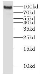 human brain tissue were subjected to SDS PAGE followed by western blot with FNab02819(EPS8 antibody) at dilution of 1:500
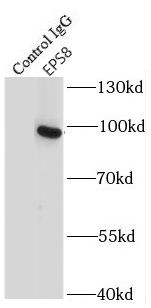 IP Result of anti-EPS8 (IP:FNab02819, 3ug; Detection:FNab02819 1:500) with HeLa cells lysate 3800ug.
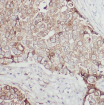 Immunohistochemistry of paraffin-embedded human breast cancer using FNab02819(EPS8 antibody) at dilution of 1:50
- Background
- Signaling adapter that controls various cellular protrusions by regulating actin cytoskeleton dynamics and architecture. Depending on its association with other signal transducers, can regulate different processes. Together with SOS1 and ABI1, forms a trimeric complex that participates in transduction of signals from Ras to Rac by activating the Rac-specific guanine nucleotide exchange factor(GEF) activity. Acts as a direct regulator of actin dynamics by binding actin filaments and has both barbed-end actin filament capping and actin bundling activities depending on the context. Displays barbed-end actin capping activity when associated with ABI1, thereby regulating actin-based motility process: capping activity is auto-inhibited and inhibition is relieved upon ABI1 interaction. Also shows actin bundling activity when associated with BAIAP2, enhancing BAIAP2-dependent membrane extensions and promoting filopodial protrusions. Involved in the regulation of processes such as axonal filopodia growth, stereocilia length, dendritic cell migration and cancer cell migration and invasion. Acts as a regulator of axonal filopodia formation in neurons: in the absence of neurotrophic factors, negatively regulates axonal filopodia formation via actin-capping activity. In contrast, it is phosphorylated in the presence of BDNF leading to inhibition of its actin-capping activity and stimulation of filopodia formation. Component of a complex with DFNB31 and MYO15A that localizes at stereocilia tips and is required for elongation of the stereocilia actin core. Indirectly involved in cell cycle progression; its degradation following ubiquitination being required during G2 phase to promote cell shape changes.