CD206 antibody

11th Anniversary Sale
Synonyms:Macrophage mannose receptor 1 (MMR)|C-type lectin domain family 13 member D|C-type lectin domain family 13 member D-like|Human mannose receptor (hMR)|Macrophage mannose receptor 1-like protein 1|MRC1|CLEC13D|CLEC13DL|MRC1L1 antibody
Catalogue No.:FNab01442Reactivity:Human, Mouse, Rat
Host:RabbitTested Application:ELISA, WB, IHC, FC
Clonality:polyclonalIsotype:IgG
Size Price
100µg Inquiry
Dispatch Time: About 3 working days
  • SPECIFICATIONS
  • CITATIONS
  • FIGURES
  • CONDITIONS
  • FAQS
Product Name
CD206 antibody
Catalogue No.
FNab01442
Size
100μg
Form
liquid
Purification
Immunogen affinity purified
Purity
≥95% as determined by SDS-PAGE
Clonality
polyclonal
Isotype
IgG
Storage
PBS with 0.02% sodium azide and 50% glycerol pH 7.3, -20℃ for 12 months(Avoid repeated freeze / thaw cycles.)
Immunogen
Immunogen
mannose receptor, C type 1
Alternative Names
Macrophage mannose receptor 1 (MMR)|C-type lectin domain family 13 member D|C-type lectin domain family 13 member D-like|Human mannose receptor (hMR)|Macrophage mannose receptor 1-like protein 1|MRC1|CLEC13D|CLEC13DL|MRC1L1 antibody
UniProt ID
P22897
Observed MW
166 kDa
Application
Tested Applications
ELISA, WB, IHC, FC
Recommended dilution
WB: 1:200-1:2000; IHC: 1:50-1:200
Validated Images
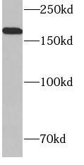
mouse kidney tissue were subjected to SDS PAGE followed by western blot with FNab01442(MRC1 antibody) at dilution of 1:500
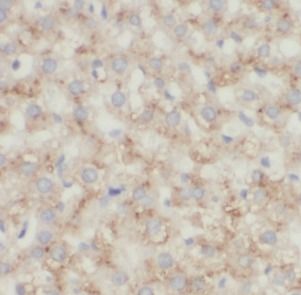
Immunohistochemistry of paraffin-embedded human liver using FNab01442(MRC1 antibody) at dilution of 1:100
Background
CD206, also named as MMR, CLEC13D and MRC1, is a type I membrane receptor that mediates the endocytosis of glycoproteins by macrophages. CD206 has been shown to bind high-mannose structures on the surface of potentially pathogenic viruses, bacteria, and fungi so that they can be neutralized by phagocytic engulfment. CD206 is a 170 kDa transmembrane protein which contains 5 domains: an amino-terminal cysteine-rich region, a fibronectin type II repeat, a series of eight tandem lectin-like carbohydrate recognition domains(responsible for the recognition of mannose and fucose), a transmembrane domain, and an intracellular carboxy-terminal tail. It is expressed on most tissue macrophages, in vitro derived dendritic cells, lymphatic and sinusoidal endothelia. This antibody recognizes the intracellular carboxy-terminal part of CD206 and MRC1L1.
IF: 4.6
Journal:
Scientific Reports
Author:
Department of Breast Surgery, First Affiliated Hospital of Kunming Medical University, Kunming, 650223, China
Cited Date:
2023-12-22
Product:
How many times can antibodies be recycled?

First, usually it's not suggested to recycle antibodies. After use, buffer system of antibodies has changed. The storage condition of recycled antibodies for different customers also varies. Thus, the performance efficiency of recycled antibodies can’t be guaranteed. Besides, FineTest ever conducted the antibody recycling assay. Assay results show recycling times of different antibodies also varies. Usually, higher antibody titer allows more repeated use. Customers can determine based on experimental requirements.

Notes: After incubation, we recycle rest antibodies to centrifuge tube and store at 4℃. High titer antibodies can be stored for a minimum of one week. Reuse about three times.

What are components of FineTest antibody buffer?

Components of FineTest antibody buffer are usually PBS with proclin300 or sodium azide, BSA, 50% glycerol. Common preservative is proclin300 or sodium azide, which is widely applied in the lab and industry.

How about the storage temperature and duration of FineTest antibodies?

Most antibodies are stored at -20℃. Directly-labeled flow cytometry antibodies should be stored at 2 - 8℃. The shelf life is one year. If after sales issues for purchased antibodies appear, return or replacement is available. Usually, antibodies can be still used after the one-year warranty. We can offer technical support services.

Is dilution required for FineTest antibodies? What’s the dilute solution?

Directly-labeled flow cytometry antibodies are ready-to-use without dilution. Other antibodies are usually concentrated. Follow the dilution ratio suggested in the manual. Dilute solution for different experiments also varies. Common antibody dilution buffers are acceptable(e.g. PBST, TBST, antibody blocking buffer).

How to retrieve antibodies for immunohistochemistry?

Common retrieval buffers: Tris-EDTA Buffer(pH 9.0); Citrate Buffer(pH 6.0)

Heat induced antibody retrieval:

Method 1: Water-bath heating: Put the beaker with retrieval buffer and slide in the boiling water bath. Keep the boiling state for 15min. Naturally cool to room temperature;

Method 2: Microwave retrieval: Put the beaker with retrieval buffer and slide in the microwave oven. Heat at high power for 5min, Switch OFF for 3min, Heat at medium power for 5min. Naturally cool to room temperature.

How to choose secondary antibodies?

(1) Secondary antibodies react with primary antibodies. Thus, secondary antibodies should be against host species of primary antibodies. E.g. If the primary antibody is derived from rabbit, the relevant secondary antibody should be against rabbit. E.g. goat anti rabbit or donkey anti rabbit.

(2) Choose secondary antibody conjugates according to the experimental type, e.g. ELISA, WB, IHC etc. Common enzyme conjugated secondary antibodies are labelled by HRP, AP etc. Fluorescin or dye labelled secondary antibodies are applied in immunofluorescence and flow cytometry(e.g. FITC, Cy3).

Product Comparison