Products
VIL1 antibody
| Size | Price |
|---|---|
| 100µg | Inquiry |
Dispatch Time:
About 3 working days
- Product Name
- VIL1 antibody
- Catalogue No.
- FNab09407
- Size
- 100μg
- Form
- liquid
- Purification
- Protein A+G purification
- Purity
- ≥95% as determined by SDS-PAGE
- Clonality
- monoclonal
- Isotype
- IgG1
- Clone ID
- 3D11
- Storage
- PBS with 0.02% sodium azide and 50% glycerol pH 7.3, -20℃ for 12 months(Avoid repeated freeze / thaw cycles.)
Immunogen
- Immunogen
- villin 1
- Alternative Names
- Villin-1|VIL1|VIL antibody
- UniProt ID
- P09327
- Observed MW
- 93 kDa
Application
- Tested Applications
- ELISA, WB, IHC, IF, IP
- Recommended dilution
- WB: 1:500-1:5000; IHC: 1:50-1:2000; IF: 1:10-1:100; IP: 1:500-1:5000
Validated Images
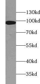 human kidney tissue were subjected to SDS PAGE followed by western blot with FNab09407(VIL1 antibody) at dilution of 1:1000
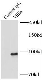 IP Result of anti-Villin (IP:FNab09407, 4ug; Detection:FNab09407 1:300) with mouse kidney tissue lysate 4000ug.
 Immunohistochemistry of paraffin-embedded human colon cancer tissue slide using FNab09407(VIL1 Antibody) at dilution of 1:50?
- Background
- Villin 1(VIL1) is a 95-kd F-actin bundling and severing protein and its expression is restricted to epithelial cells with a brush border, like epithelial cells of the intestinal mucosa, gall bladder, renal proximal tubules and ductuli efferentes of the testis. VIL1 has been reported to be an epithelial cell-specific anti-apoptotic protein, and to have an important function in regulating actin dynamics, cell morphology, epithelial-to-mesenchymal transitions, cell migration and cell survival. In addition, VIL1 is a useful diagnostic marker for of various cancer, like cervical and endometrial adenocarcinomas, renal cell carcinoma. VIL1 was recently identified as a novel biomarker predictive for postoperative recurrence and poorer prognosis of high serum AFP associated HCC.