Products
MAPK12 antibody
| Size | Price |
|---|---|
| 100µg | Inquiry |
Dispatch Time:
About 3 working days
- Product Name
- MAPK12 antibody
- Catalogue No.
- FNab04989
- Size
- 100μg
- Form
- liquid
- Purification
- Immunogen affinity purified
- Purity
- ≥95% as determined by SDS-PAGE
- Clonality
- polyclonal
- Isotype
- IgG
- Storage
- PBS with 0.02% sodium azide and 50% glycerol pH 7.3, -20℃ for 12 months(Avoid repeated freeze / thaw cycles.)
Immunogen
- Immunogen
- mitogen-activated protein kinase 12
- Alternative Names
- Mitogen-activated protein kinase 12 (MAP kinase 12 antibody, MAPK 12)|Extracellular signal-regulated kinase 6 (ERK-6)|Mitogen-activated protein kinase p38 gamma (MAP kinase p38 gamma)|Stress-activated protein kinase 3|MAPK12|ERK6|SAPK3 antibody
- UniProt ID
- P53778
- Observed MW
- 42 kDa
Application
- Tested Applications
- ELISA, WB, IHC
- Recommended dilution
- WB: 1:500-1:2000; IHC: 1:20-1:200
Validated Images
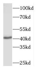 human heart tissue were subjected to SDS PAGE followed by western blot with FNab04989(MAPK12 antibody) at dilution of 1:500
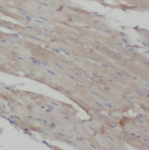 Immunohistochemistry of paraffin-embedded human heart using FNab04989(MAPK12 antibody) at dilution of 1:100
- Background
- Serine/threonine kinase which acts as an essential component of the MAP kinase signal transduction pathway. MAPK12 is one of the four p38 MAPKs which play an important role in the cascades of cellular responses evoked by extracellular stimuli such as proinflammatory cytokines or physical stress leading to direct activation of transcription factors such as ELK1 and ATF2. Accordingly, p38 MAPKs phosphorylate a broad range of proteins and it has been estimated that they may have approximately 200 to 300 substrates each. Some of the targets are downstream kinases such as MAPKAPK2, which are activated through phosphorylation and further phosphorylate additional targets. Plays a role in myoblast differentiation and also in the down-regulation of cyclin D1 in response to hypoxia in adrenal cells suggesting MAPK12 may inhibit cell proliferation while promoting differentiation. Phosphorylates DLG1. Following osmotic shock, MAPK12 in the cell nucleus increases its association with nuclear DLG1, thereby causing dissociation of DLG1-SFPQ complexes. This function is independent of its catalytic activity and could affect mRNA processing and/or gene transcription to aid cell adaptation to osmolarity changes in the environment. Regulates UV-induced checkpoint signaling and repair of UV-induced DNA damage and G2 arrest after gamma-radiation exposure. MAPK12 is involved in the regulation of SLC2A1 expression and basal glucose uptake in L6 myotubes; and negatively regulates SLC2A4 expression and contraction-mediated glucose uptake in adult skeletal muscle. C-Jun(JUN) phosphorylation is stimulated by MAPK14 and inhibited by MAPK12, leading to a distinct AP-1 regulation. MAPK12 is required for the normal kinetochore localization of PLK1, prevents chromosomal instability and supports mitotic cell viability. MAPK12-signaling is also positively regulating the expansion of transient amplifying myogenic precursor cells during muscle growth and regeneration.