Products
CEP290 antibody
Research Area:
| Size | Price |
|---|---|
| 100µg | Inquiry |
Dispatch Time:
About 3 working days
- Product Name
- CEP290 antibody
- Catalogue No.
- FNab01602
- Size
- 100μg
- Form
- liquid
- Purification
- Immunogen affinity purified
- Purity
- ≥95% as determined by SDS-PAGE
- Clonality
- polyclonal
- Isotype
- IgG
- Storage
- PBS with 0.02% sodium azide and 50% glycerol pH 7.3, -20℃ for 12 months(Avoid repeated freeze / thaw cycles.)
Immunogen
- Immunogen
- centrosomal protein 290kDa
- Alternative Names
- Centrosomal protein of 290 kDa (Cep290)|Bardet-Biedl syndrome 14 protein|Cancer/testis antigen 87 (CT87)|Nephrocystin-6|Tumor antigen se2-2|CEP290|BBS14|KIAA0373|NPHP6 antibody
- UniProt ID
- O15078
- Observed MW
- 290 kDa, 150-180 kDa, 80 kDa, 60 kDa
Application
- Tested Applications
- ELISA, WB, IP, IHC, IF
- Recommended dilution
- WB: 1:200-1:2000; IP: 1:500-1:5000; IHC: 1:20-1:200; IF: 1:50-1:500
Validated Images
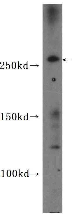 HeLa cells were subjected to SDS PAGE followed by western blot with FNab01602(CEP290 Antibody) at dilution of 1:600
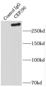 IP result of anti-CEP290(FNab01602 for IP and Detection)with HeLa cells.
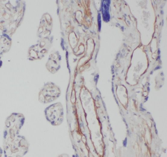 Immunohistochemistry of paraffin-embedded human placenta using FNab01602(CEP290 antibody) at dilution of 1:50
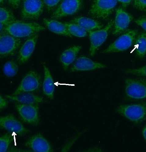 Immunofluorescent analysis of ( -20℃ Ethanol ) fixed HeLa cells using FNab01602 ( CEP290 Antibody) at dilution of 1:50 and Alexa Fluor 488-conjugated AffiniPure Goat Anti-Rabbit IgG(H+L)
- Background
- Studies in the Chlamydomonas model system has shown that the ciliary protein CEP290 is a critical component of these Y-link junctions. Additionally, numerous studies have demonstrated that CEP290’s function is critical for IFT—in CEP290 knockdown experiments, many proteins that would normally localize to the cilium fail to do so, and cilium formation is disrupted or absent. Mutations in CEP290 are accountable for cases of nephronophthisis, Leber congenital amaurosis and Joubert syndrome. In IF analtsis of HeLa cells, the white arrows show centrosome and cilium staining.