CTNNB1 antibody

Synonyms:Catenin beta-1|Beta-catenin|CTNNB1|CTNNB antibody
Catalogue No.:FNab00882Reactivity:Human, Mouse, Rat, Pig
Host:RabbitTested Application:ELISA, IF, WB, IHC, IP
Clonality:polyclonalIsotype:IgG
Size Price
100µg Inquiry
Dispatch Time: About 3 working days
  • SPECIFICATIONS
  • CITATIONS
  • FIGURES
  • CONDITIONS
  • FAQS
Product Name
CTNNB1 antibody
Catalogue No.
FNab00882
Size
100μg
Form
liquid
Purification
Immunogen affinity purified
Purity
≥95% as determined by SDS-PAGE
Clonality
polyclonal
Isotype
IgG
Storage
PBS with 0.02% sodium azide and 50% glycerol pH 7.3, -20℃ for 12 months(Avoid repeated freeze / thaw cycles.)
Immunogen
Immunogen
catenin(cadherin-associated protein), beta 1, 88kDa
Alternative Names
Catenin beta-1|Beta-catenin|CTNNB1|CTNNB antibody
UniProt ID
P35222
Observed MW
90 kDa
Application
Tested Applications
ELISA, IF, WB, IHC, IP
Recommended dilution
WB: 1:1000-1:4000; IP: 1:500-1:1000; IHC: 1:100-1:400; IF: 1:20-1:200
Validated Images
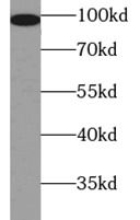
mouse lung tissue were subjected to SDS PAGE followed by western blot with FNab00882(b-catentin antibody) at dilution of 1:1000
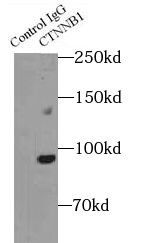
IP Result of anti-beta-Catenin (IP: FNab00882, 4ug; Detection: FNab00882 1:500) with mouse liver tissue lysate 3200ug.
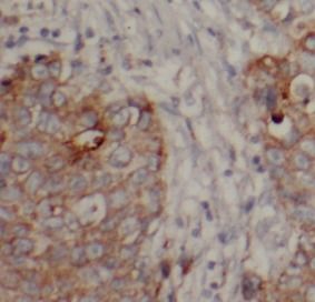
Immunohistochemistry of paraffin-embedded human breast cancer tissue slide using FNab00882(B-catenin Antibody) at dilution of 1:200
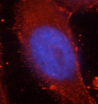
Immunofluorescent analysis of HepG2 cells, using FNab00882(B-catenin Antibody) at 1:50 dilution and Rhodamine-labeled goat anti-rabbit IgG (red). Blue pseudocolor = DAPI (fluorescent DNA dye).
Background
The protein encoded by this gene is part of a complex of proteins that constitute adherens junctions (AJs). AJs are necessary for the creation and maintenance of epithelial cell layers by regulating cell growth and adhesion between cells. The encoded protein also anchors the actin cytoskeleton and may be responsible for transmitting the contact inhibition signal that causes cells to stop dividing once the epithelial sheet is complete. Finally, this protein binds to the product of the APC gene, which is mutated in adenomatous polyposis of the colon. Mutations in this gene are a cause of colorectal cancer (CRC), pilomatrixoma (PTR), medulloblastoma (MDB), and ovarian cancer. Alternative splicing results in multiple transcript variants.
IF: 2.552
Journal:
Cardiovascular Diagnosis and Therapy
Author:
Cardiovascular Department, The First Affiliated Hospital of Anhui Medical University, Hefei, China; 2Cardiovascular Department for Gerontism, The Second Affiliated Hospital of Anhui Medical University, Hefei, China
Cited Date:
2023-06-16
Product:
How many times can antibodies be recycled?

First, usually it's not suggested to recycle antibodies. After use, buffer system of antibodies has changed. The storage condition of recycled antibodies for different customers also varies. Thus, the performance efficiency of recycled antibodies can’t be guaranteed. Besides, FineTest ever conducted the antibody recycling assay. Assay results show recycling times of different antibodies also varies. Usually, higher antibody titer allows more repeated use. Customers can determine based on experimental requirements.

Notes: After incubation, we recycle rest antibodies to centrifuge tube and store at 4℃. High titer antibodies can be stored for a minimum of one week. Reuse about three times.

What are components of FineTest antibody buffer?

Components of FineTest antibody buffer are usually PBS with proclin300 or sodium azide, BSA, 50% glycerol. Common preservative is proclin300 or sodium azide, which is widely applied in the lab and industry.

How about the storage temperature and duration of FineTest antibodies?

Most antibodies are stored at -20℃. Directly-labeled flow cytometry antibodies should be stored at 2 - 8℃. The shelf life is one year. If after sales issues for purchased antibodies appear, return or replacement is available. Usually, antibodies can be still used after the one-year warranty. We can offer technical support services.

Is dilution required for FineTest antibodies? What’s the dilute solution?

Directly-labeled flow cytometry antibodies are ready-to-use without dilution. Other antibodies are usually concentrated. Follow the dilution ratio suggested in the manual. Dilute solution for different experiments also varies. Common antibody dilution buffers are acceptable(e.g. PBST, TBST, antibody blocking buffer).

How to retrieve antibodies for immunohistochemistry?

Common retrieval buffers: Tris-EDTA Buffer(pH 9.0); Citrate Buffer(pH 6.0)

Heat induced antibody retrieval:

Method 1: Water-bath heating: Put the beaker with retrieval buffer and slide in the boiling water bath. Keep the boiling state for 15min. Naturally cool to room temperature;

Method 2: Microwave retrieval: Put the beaker with retrieval buffer and slide in the microwave oven. Heat at high power for 5min, Switch OFF for 3min, Heat at medium power for 5min. Naturally cool to room temperature.

How to choose secondary antibodies?

(1) Secondary antibodies react with primary antibodies. Thus, secondary antibodies should be against host species of primary antibodies. E.g. If the primary antibody is derived from rabbit, the relevant secondary antibody should be against rabbit. E.g. goat anti rabbit or donkey anti rabbit.

(2) Choose secondary antibody conjugates according to the experimental type, e.g. ELISA, WB, IHC etc. Common enzyme conjugated secondary antibodies are labelled by HRP, AP etc. Fluorescin or dye labelled secondary antibodies are applied in immunofluorescence and flow cytometry(e.g. FITC, Cy3).

Product Comparison