Abstract: Exosome was early found in the supernatant of sheep erythrocyte cultured in vitro. It's clearly defined as the extracellular vesicle and plays an important role in cell communication and tissue microenvironment regulation. After isolation, characterization methods are required to confirm whether the isolated target is exosome.
Keywords: Exosome Characterization, Exosome Isolation, Exosome Marker Antibodies
1. Exosome Structure
Exosomes are released by many cells and exist in various body fluid, such as B cell, dendritic cell, T cell, epidermal cell, platelet, amniotic fluid, ascite, saliva, nasal lavage fluid, serum, plasma, milk, urine, cerebrospinal fluid etc. It's estimated that the exosome generation in cells may physiologically remove redundant and unnecessary components to maintain intracellular homeostasis.
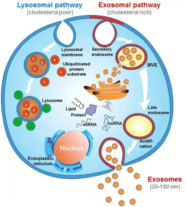
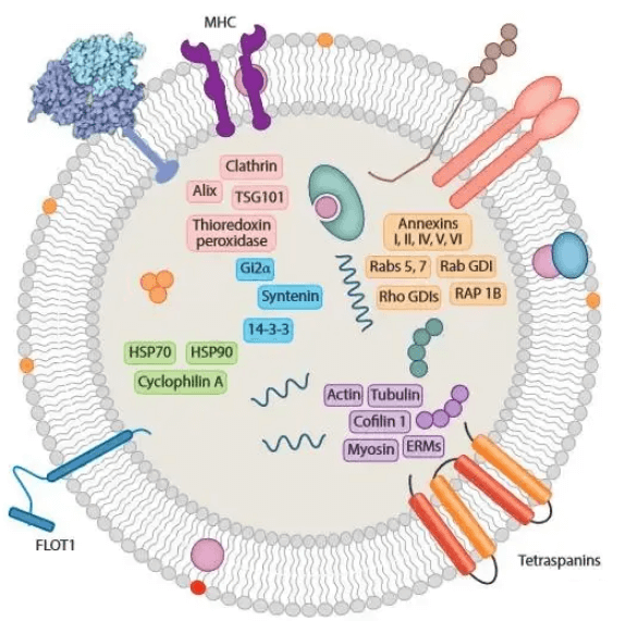
Exosomes with lipid bilayer structure are rich in cholesterol and sphingomyelin. Nearly all exosomes have tubulin, actin-binding protein and tetraspanin. Components of exosomes derived from different cells are also distinct. (e.g. Intestinal epithelial cell derived exosomes contain various metabolic enzymes and intestinal tissue specific A33 antigen; B cell derived exosomes are rich in CD86 and MHC molecule; Pro-apoptotic FasL receptor is on the surface of T cell derived exosomes. ) The typical exosome structure is shown below.
2. Exosomes in Cancer
Exosomes are related to immune reaction, viral pathogenicity, pregnancy, cardiovascular diseases, CNS diseases and oncology progress etc. Proteins, metabolites, and nucleic acids delivered by exosomes to receptor cells efficiently change their biological response. The exosome mediated response may be disease promotion or suppression. Exosomes promote the potential effect of treatment and control for many diseases(e.g. neurodegenerative disease and cancer) during regulating intrinsic properties of complex intracellular pathway.
3. Exosome Storage Conditions
The extracted exosome is usually suspended in phosphate buffer. Cryopreservation is commonly used but may change the shape and physical property. Multiple layer vesicles may also form and aggregate. Freeze-thaw cycle will change biological features, content and marker composition of surface molecules on exosomes. It's suggested that freshly isolated exosome should be immediately analyzed or shortly stored at 4℃/-20℃. -80℃ is more suitable for clinical treatment and long storage.
4. Exosome Characterization Methods
Exosome characterization methods are involved in many aspects, e.g. physical characteristics, surface molecular marker etc.
Transmission Electron Microscope(TEM): Observe ultrastructural morphology of bi-layered exosome properly (saucer or cup-shaped).
Nanoparticle Tracking Analysis(NTA): ensure the original state exosome. Fast detection to offer
Western Blot Analysis: Exosome marker proteins include tetraspanin(e.g. CD9, CD63 and CD81), cytoplasmic protein(e.g. actin, annexins etc.), molecule involved in biologic function (e.g. ALIX, TSG101, HSP70, HSP90), and specific protein secreted by cells.
Lipophilic Dye Labelling: In the published articles, most exosomes are labelled by lipophilic dye in vivo and vitro. There are two types of lipophilic dyes: PKH67(green fluorescence) / PKH26(red fluorescence). Since they can bind with bilayer lipid membrane steadily, the good staining effect and wide applications are possible.
5. Recommended Antibodies
|
Catalogue No. |
Product Name |
Type |
Reactivity |
Applications |
|
anti- CD9 antibody |
Rabbit pAb |
Human, Rat, Mouse |
ELISA, WB, IHC, FC |
|
|
anti- CD9 antibody |
Mouse mAb |
Human |
ELISA, WB, IHC, FC |
|
|
anti- CD63 antibody |
Rabbit pAb |
Human, Rat, Mouse |
ELISA, IHC, WB |
|
|
anti- CD81 antibody |
Rabbit pAb |
Human, Rat, Mouse |
ELISA, IHC, WB |
|
|
anti- CD81 antibody |
Rabbit pAb |
Human, Rat, Mouse |
ELISA, IHC, WB |
|
|
anti- PDCD6IP antibody |
Rabbit pAb |
Human, Rat, Mouse |
ELISA, WB, IHC |
|
|
anti- TSG101 antibody |
Rabbit pAb |
Human, Rat, Mouse |
ELISA, WB, IHC |
|
|
anti- TSG101 antibody |
Mouse mAb |
Human, Rat, Mouse |
ELISA, WB, IHC,FC |
|
|
anti- HSP70 antibody |
Rabbit pAb |
Human, Rat, Mouse |
ELISA, IHC, WB, IF, FC, IP |
|
|
anti- HSP70 antibody |
Rabbit pAb |
Human, Rat, Mouse |
ELISA, WB,IHC |
|
|
anti- HSP70 antibody |
Mouse mAb |
Human, Rat, Mouse |
ELISA, WB,IHC |
|
|
anti- HSP90 antibody |
Rabbit pAb |
Human, Rat, Mouse |
ELISA, IF, IHC, IP, WB, FC |
|
|
anti- HSP90 antibody |
Mouse mAb |
Human, Rat, Mouse |
ELISA, WB, IHC, FC |
REFERENCES
[1]Exosome Processing and Characterization Approaches for Research and Technology Development, PMID: 35332686.
[2]Isolation and characterization of exosomes for cancer research, PMID: 33168028.