Products
PE-Cy5.5 Anti-Mouse CD161/NK1.1 Antibody(PK136)
Inquiry
Dispatch Time:
About 3 working days
- Product Name
- PE-Cy5.5 Anti-Mouse CD161/NK1.1 Antibody(PK136)
- Catalogue No.
- PC55-30029
- Form
- liquid
- Conjugation
- PE-Cy5.5
- Conjugation Information
- PE-Cy5.5 is designed to be excited by the Blue (488 nm), Green (532 nm) and yellow-green (561 nm) lasers and detected using an optical filter centered near 690 nm (e.g., a 690/50 nm bandpass filter).
- Clonality
- Monoclonal
- Isotype
- IgG2a, κ
- Clone ID
- PK136
- Storage
- PBS with 0. 1% sodium azide, 1%BSA, pH 7.3, 2-8℃ for 12 months (Avoid repeated freeze / thaw cycles.)
Immunogen
Application
- Tested Applications
- FC
- Recommended dilution
- Volume per test: 5μL. Each lot of this antibody is quality control tested by flow cytometric analysis. The amount of the reagent is suggested to be used 5 µL of antibody per test (million cells in 100 µL staining volume or per 100 µL of whole blood). Please check your vial before the experiment. Since applications vary, the appropriate dilutions must be determined for individual use.
Validated Images
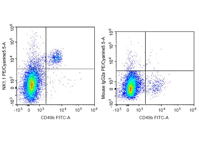 C57BL/6 murine splenocytes are stained with FITC Anti-Mouse CD49b Antibody and PE-Cy5.5 Anti-Mouse CD161/NK1.1 Antibody (Left). Splenocytes are stained with FITC Anti-Mouse CD49b Antibody and PE-Cy5.5 Mouse IgG2a, κ Isotype Control (Right).
- Background
- NK-1.1 surface antigen, also known as CD161b/CD161c and Ly-55, is encoded by the NKR-P1B/NKR-P1C gene. It is expressed on NK cells and NK-T cells in some mouse strains, including C57BL/6, FVB/N, and NZB, but not AKR, BALB/c, CBA/J, C3H, DBA/1, DBA/2, NOD, SJL, and 129. Expression of NKR-P1C antigen has been correlated with lysis of tumor cells in vitro and rejection of bone marrow allografts in vivo. NK-1.1 has also been shown to play a role in NK cell activation, IFN-γ production, and cytotoxic granule release. NK-1.1 and DX5 are commonly used as mouse NK cell markers.