Products
PE-FineTest®594 Anti-Mouse CD146 Antibody(ME-9F1)
Inquiry
Dispatch Time:
About 3 working days
- Product Name
- PE-FineTest®594 Anti-Mouse CD146 Antibody(ME-9F1)
- Catalogue No.
- PE5-30197
- Form
- liquid
- Conjugation
- PE-FineTest®594
- Conjugation Information
- PE-FineTest®594 is designed to be excited by the blue (488 nm), Green (532 nm) and yellow-green (561 nm) lasers and detected using an optical filter centered near 620 nm (e.g., a 610/20 nm bandpass filter).
- Clonality
- Monoclonal
- Isotype
- IgG2a, κ
- Clone ID
- ME-9F1
- Storage
- PBS with 0. 1% sodium azide, 1%BSA, pH 7.3, 2-8℃ for 12 months (Avoid repeated freeze / thaw cycles.)
Immunogen
- Alternative Names
- S-Endo 1 antigen|MUC18|MCAM|Mel-CAM|A32 antigen antibody
- UniProt ID
- Q8R2Y2
Application
- Tested Applications
- FC
- Recommended dilution
- Volume per test: 5μL. Each lot of this antibody is quality control tested by flow cytometric analysis. The amount of the reagent is suggested to be used 5 µL of antibody per test (million cells in 100 µL staining volume or per 100 µL of whole blood). Please check your vial before the experiment. Since applications vary, the appropriate dilutions must be determined for individual use.
Validated Images
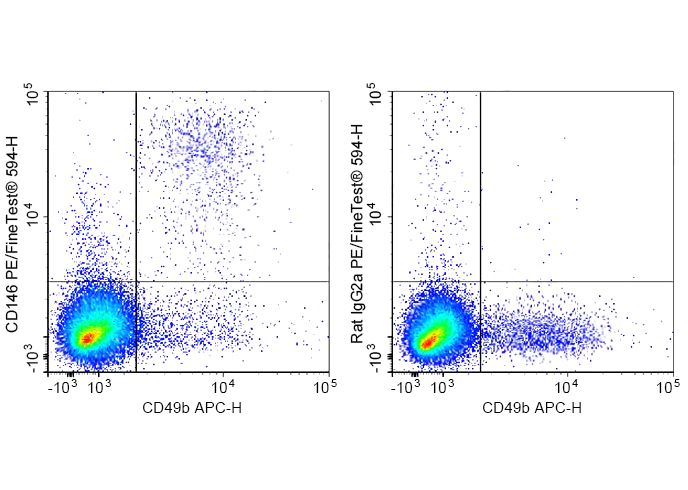 Staining of C57BL/6 murine splenocytes cells with APC Anti-Mouse CD49b Antibody and PE-FineTest®594 Anti-Mouse NKG2A/C/E Antibody(ME-9F1) (left) or PE-FineTest®594 Rat IgG2a,κ Isotype Control (right). Total viable cells were used for analysis.
- Background
- CD146, also known as melanoma cell adhesion molecule (MCAM or Mel-CAM), MUC18, S-Endo1, and A32 antigen, is an integral membrane glycoprotein that belongs to the Ig superfamily. CD146 is strongly expressed by murine vascular endothelial cells. It is expressed on about 30% of neutrophils and 60% of NK cells. Unlike in humans, CD146 is undetectable on monocytes, dendritic cells, T cells, NKT cells, B cells, or smooth muscle cells in mouse. It has been reported that an increase in CD146 expression is associated with NK cell maturation. Combined with using CD27 and CD11b staining, CD146 may be an alternative marker to detect final stages of NK cell maturation and define NK cell subsets. CD146+ NK cells were found to be less cytotoxic and to produce less IFNγ than CD146- NK cells upon stimulation with target cells or activating antibodies. The role of CD146 on NK cell migration has yet to be investigated. The identification of CD146 ligand(s) will be crucial to address this issue.