Products
KLRK1 antibody
| Size | Price |
|---|---|
| 100ug | Inquiry |
Dispatch Time:
About 3 working days
- Product Name
- KLRK1 antibody
- Catalogue No.
- FNab10477
- Size
- 100μg
- Form
- liquid
- Purification
- Immunogen affinity purified
- Purity
- ≥95% as determined by SDS-PAGE
- Clonality
- polyclonal
- Isotype
- IgG
- Storage
- PBS with 0.02% sodium azide and 50% glycerol pH 7.3, -20℃ for 12 months(Avoid repeated freeze / thaw cycles.)
Immunogen
- Immunogen
- NKG2-D type II integral membrane protein
- Alternative Names
- NKG2-D type II integral membrane protein|Killer cell lectin-like receptor subfamily K member 1|NK cell receptor D|NKG2-D-activating NK receptor|KLRK1|D12S2489E|NKG2D antibody
- UniProt ID
- P26718
- Observed MW
- 30 kDa
Application
- Tested Applications
- ELISA, WB
- Recommended dilution
- WB: 1:500-1:2000
Validated Images
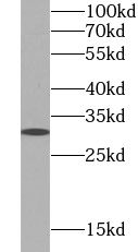 HepG2 cells were subjected to SDS PAGE followed by western blot with FNab10477(KLRK1 Antibody) at dilution of 1:1000
- Background
- Natural killer (NK) cells are lymphocytes that can mediate lysis of certain tumor cells and virus-infected cells without previous activation. They can also regulate specific humoral and cell-mediated immunity. NK cells preferentially express several calcium-dependent (C-type) lectins, which have been implicated in the regulation of NK cell function. The NKG2 gene family is located within the NK complex, a region that contains several C-type lectin genes preferentially expressed in NK cells. This gene encodes a member of the NKG2 family. The encoded transmembrane protein is characterized by a type II membrane orientation (has an extracellular C terminus) and the presence of a C-type lectin domain. It binds to a diverse family of ligands that include MHC class I chain-related A and B proteins and UL-16 binding proteins, where ligand-receptor interactions can result in the activation of NK and T cells. The surface expression of these ligands is important for the recognition of stressed cells by the immune system, and thus this protein and its ligands are therapeutic targets for the treatment of immune diseases and cancers. Read-through transcription exists between this gene and the upstream KLRC4 (killer cell lectin-like receptor subfamily C, member 4) family member in the same cluster.