Products
ABCA4 antibody
| Size | Price |
|---|---|
| 100µg | Inquiry |
Dispatch Time:
About 3 working days
- Product Name
- ABCA4 antibody
- Catalogue No.
- FNab09922
- Size
- 100μg
- Form
- liquid
- Purification
- Immunogen affinity purified
- Purity
- ≥95% as determined by SDS-PAGE
- Clonality
- polyclonal
- Isotype
- IgG
- Storage
- PBS with 0.02% sodium azide and 50% glycerol pH 7.3, -20℃ for 12 months(Avoid repeated freeze / thaw cycles.)
Immunogen
- Immunogen
- ABCA4
- Alternative Names
- Retinal-specific phospholipid-transporting ATPase ABCA4|ATP-binding cassette sub-family A member 4|RIM ABC transporter (RIM proteinv antibody, RmP)|Retinal-specific ATP-binding cassette transporter|Stargardt disease protein|ABCA4|ABCR antibody
- UniProt ID
- P78363
- Observed MW
- 256 kDa
Application
- Tested Applications
- ELISA, WB, IF
- Recommended dilution
- WB: 1:500 - 1:2000; IF: 1:50 - 1:200
Validated Images
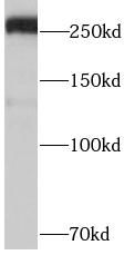 BT474 cells were subjected to SDS PAGE followed by western blot with FNab09922 (ABCA4 antibody) at dilution of 1:1000
- Background
- The membrane-associated protein encoded by this gene is a member of the superfamily of ATP-binding cassette (ABC) transporters. ABC proteins transport various molecules across extra- and intracellular membranes. ABC genes are divided into seven distinct subfamilies (ABC1, MDR/TAP, MRP, ALD, OABP, GCN20, White). This protein is a member of the ABC1 subfamily. Members of the ABC1 subfamily comprise the only major ABC subfamily found exclusively in multicellular eukaryotes. This protein is a retina-specific ABC transporter with N-retinylidene-PE as a substrate. It is expressed exclusively in retina photoreceptor cells, indicating the gene product mediates transport of an essental molecule across the photoreceptor cell membrane. Mutations in this gene are found in patients diagnosed with Stargardt disease, a form of juvenile-onset macular degeneration. Mutations in this gene are also associated with retinitis pigmentosa-19, cone-rod dystrophy type 3, early-onset severe retinal dystrophy, fundus flavimaculatus, and macular degeneration age-related 2.