Products
VCP antibody
| Size | Price |
|---|---|
| 100µg | Inquiry |
Dispatch Time:
About 3 working days
- Product Name
- VCP antibody
- Catalogue No.
- FNab09382
- Size
- 100μg
- Form
- liquid
- Purification
- Protein A+G purification
- Purity
- ≥95% as determined by SDS-PAGE
- Clonality
- monoclonal
- Isotype
- IgG1
- Clone ID
- 7B11
- Storage
- PBS with 0.02% sodium azide and 50% glycerol pH 7.3, -20℃ for 12 months(Avoid repeated freeze / thaw cycles.)
Immunogen
- Immunogen
- valosin-containing protein
- Alternative Names
- Transitional endoplasmic reticulum ATPase (TER ATPase)|15S Mg(2+)-ATPase p97 subunit|Valosin-containing protein (VCP)|VCP|HEL-220|HEL-S-70 antibody
- UniProt ID
- P55072
- Observed MW
- 89 kDa
Application
- Tested Applications
- ELISA, WB, IHC, IF
- Recommended dilution
- WB: 1:500-1:5000; IHC: 1:20-1:200;IF: 1:20-1:200
Validated Images
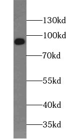 RAW 264.7 cells were subjected to SDS PAGE followed by western blot with FNab09382(VCP Antibody) at dilution of 1:1000
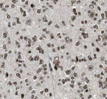 Immunohistochemistry of paraffin-embedded human gliomas tissue slide using FNab09382(VCP Antibody) at dilution of 1:50
- Background
- Necessary for the fragmentation of Golgi stacks during mitosis and for their reassembly after mitosis. Involved in the formation of the transitional endoplasmic reticulum(tER). The transfer of membranes from the endoplasmic reticulum to the Golgi apparatus occurs via 50-70 nm transition vesicles which derive from part-rough, part-smooth transitional elements of the endoplasmic reticulum(tER). Vesicle budding from the tER is an ATP-dependent process. The ternary complex containing UFD1L, VCP and NPLOC4 binds ubiquitinated proteins and is necessary for the export of misfolded proteins from the ER to the cytoplasm, where they are degraded by the proteasome. The NPLOC4-UFD1L-VCP complex regulates spindle disassembly at the end of mitosis and is necessary for the formation of a closed nuclear envelope. Regulates E3 ubiquitin-protein ligase activity of RNF19A. Component of the VCP/p97-AMFR/gp78 complex that participates in the final step of the sterol-mediated ubiquitination and endoplasmic reticulum-associated degradation(ERAD) of HMGCR. Also involved in DNA damage response: recruited to double-strand breaks(DSBs) sites in a RNF8-and RNF168-dependent manner and promotes the recruitment of TP53BP1 at DNA damage sites. Recruited to stalled replication forks by SPRTN: may act by mediating extraction of DNA polymerase eta(POLH) to prevent excessive translesion DNA synthesis and limit the incidence of mutations induced by DNA damage. Required for cytoplasmic retrotranslocation of stressed/damaged mitochondrial outer-membrane proteins and their subsequent proteasomal degradation. Essential for the maturation of ubiquitin-containing autophagosomes and the clearance of ubiquitinated protein by autophagy(PubMed:20104022).