TXK antibody

Synonyms:Tyrosine-protein kinase TXK|Protein-tyrosine kinase 4|Resting lymphocyte kinase|TXK|PTK4|RLK antibody
Catalogue No.:FNab09121Reactivity:Human
Host:RabbitTested Application:ELISA, IHC, IF
Clonality:polyclonalIsotype:IgG
Size Price
100µg Inquiry
Dispatch Time: About 3 working days
  • SPECIFICATIONS
  • FIGURES
  • CONDITIONS
  • FAQS
Product Name
TXK antibody
Catalogue No.
FNab09121
Size
100μg
Form
liquid
Purification
Immunogen affinity purified
Purity
≥95% as determined by SDS-PAGE
Clonality
polyclonal
Isotype
IgG
Storage
PBS with 0.02% sodium azide and 50% glycerol pH 7.3, -20℃ for 12 months(Avoid repeated freeze / thaw cycles.)
Immunogen
Immunogen
TXK tyrosine kinase
Alternative Names
Tyrosine-protein kinase TXK|Protein-tyrosine kinase 4|Resting lymphocyte kinase|TXK|PTK4|RLK antibody
UniProt ID
P42681
Observed MW
61 kDa
Application
Tested Applications
ELISA, IHC, IF
Recommended dilution
IF: 1:10-1:100; IHC: 1:20-1:200
Validated Images
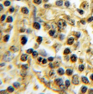
Immunohistochemical of paraffin-embedded human lymphoma using FNab09121(TXK antibody) at dilution of 1:100
Background
Non-receptor tyrosine kinase that plays a redundant role with ITK in regulation of the adaptive immune response. Regulates the development, function and differentiation of conventional T-cells and nonconventional NKT-cells. When antigen presenting cells(APC) activate T-cell receptor(TCR), a series of phosphorylation lead to the recruitment of TXK to the cell membrane, where it is phosphorylated at Tyr-420. Phosphorylation leads to TXK full activation. Contributes also to signaling from many receptors and participates in multiple downstream pathways, including regulation of the actin cytoskeleton. Like ITK, can phosphorylate PLCG1, leading to its localization in lipid rafts and activation, followed by subsequent cleavage of its substrates. In turn, the endoplasmic reticulum releases calcium in the cytoplasm and the nuclear activator of activated T-cells(NFAT) translocates into the nucleus to perform its transcriptional duty. With PARP1 and EEF1A1, TXK forms a complex that acts as a T-helper 1(Th1) cell-specific transcription factor and binds the promoter of IFNG to directly regulate its transcription, and is thus involved importantly in Th1 cytokine production. Phosphorylates both PARP1 and EEF1A1. Phosphorylates also key sites in LCP2 leading to the up-regulation of Th1 preferred cytokine IL-2. Phosphorylates 'Tyr-201' of CTLA4 which leads to the association of PI-3 kinase with the CTLA4 receptor.
How many times can antibodies be recycled?

First, usually it's not suggested to recycle antibodies. After use, buffer system of antibodies has changed. The storage condition of recycled antibodies for different customers also varies. Thus, the performance efficiency of recycled antibodies can’t be guaranteed. Besides, FineTest ever conducted the antibody recycling assay. Assay results show recycling times of different antibodies also varies. Usually, higher antibody titer allows more repeated use. Customers can determine based on experimental requirements.

Notes: After incubation, we recycle rest antibodies to centrifuge tube and store at 4℃. High titer antibodies can be stored for a minimum of one week. Reuse about three times.

What are components of FineTest antibody buffer?

Components of FineTest antibody buffer are usually PBS with proclin300 or sodium azide, BSA, 50% glycerol. Common preservative is proclin300 or sodium azide, which is widely applied in the lab and industry.

How about the storage temperature and duration of FineTest antibodies?

Most antibodies are stored at -20℃. Directly-labeled flow cytometry antibodies should be stored at 2 - 8℃. The shelf life is one year. If after sales issues for purchased antibodies appear, return or replacement is available. Usually, antibodies can be still used after the one-year warranty. We can offer technical support services.

Is dilution required for FineTest antibodies? What’s the dilute solution?

Directly-labeled flow cytometry antibodies are ready-to-use without dilution. Other antibodies are usually concentrated. Follow the dilution ratio suggested in the manual. Dilute solution for different experiments also varies. Common antibody dilution buffers are acceptable(e.g. PBST, TBST, antibody blocking buffer).

How to retrieve antibodies for immunohistochemistry?

Common retrieval buffers: Tris-EDTA Buffer(pH 9.0); Citrate Buffer(pH 6.0)

Heat induced antibody retrieval:

Method 1: Water-bath heating: Put the beaker with retrieval buffer and slide in the boiling water bath. Keep the boiling state for 15min. Naturally cool to room temperature;

Method 2: Microwave retrieval: Put the beaker with retrieval buffer and slide in the microwave oven. Heat at high power for 5min, Switch OFF for 3min, Heat at medium power for 5min. Naturally cool to room temperature.

How to choose secondary antibodies?

(1) Secondary antibodies react with primary antibodies. Thus, secondary antibodies should be against host species of primary antibodies. E.g. If the primary antibody is derived from rabbit, the relevant secondary antibody should be against rabbit. E.g. goat anti rabbit or donkey anti rabbit.

(2) Choose secondary antibody conjugates according to the experimental type, e.g. ELISA, WB, IHC etc. Common enzyme conjugated secondary antibodies are labelled by HRP, AP etc. Fluorescin or dye labelled secondary antibodies are applied in immunofluorescence and flow cytometry(e.g. FITC, Cy3).

Product Comparison