Products
SOCS3 antibody
Research Area:
| Size | Price |
|---|---|
| 100µg | Inquiry |
Dispatch Time:
About 3 working days
- Product Name
- SOCS3 antibody
- Catalogue No.
- FNab08100
- Size
- 100μg
- Form
- liquid
- Purification
- Immunogen affinity purified
- Purity
- ≥95% as determined by SDS-PAGE
- Clonality
- polyclonal
- Isotype
- IgG
- Storage
- PBS with 0.02% sodium azide and 50% glycerol pH 7.3, -20℃ for 12 months(Avoid repeated freeze / thaw cycles.)
Immunogen
- Immunogen
- suppressor of cytokine signaling 3
- Alternative Names
- Suppressor of cytokine signaling 3 (SOCS-3)|Cytokine-inducible SH2 protein 3 (CIS-3)|STAT-induced STAT inhibitor 3 (SSI-3)|SOCS3|CIS3|SSI3 antibody
- UniProt ID
- O14543
- Observed MW
- 30 kDa
Application
- Tested Applications
- ELISA, IHC, WB, IF, IP
- Recommended dilution
- WB: 1:200-1:2000; IP: 1:200-1:2000; IHC: 1:20-1:200; IF: 1:10-1:100
Validated Images
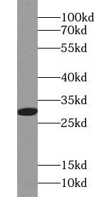 K-562 cells were subjected to SDS PAGE followed by western blot with FNab08100(SOCS3 antibody) at dilution of 1:500
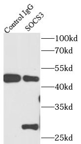 IP Result of anti-SOCS3 (IP:FNab08100,4ug; Detection: FNab08100 1:500) with K-562 cells lysate 1720ug.
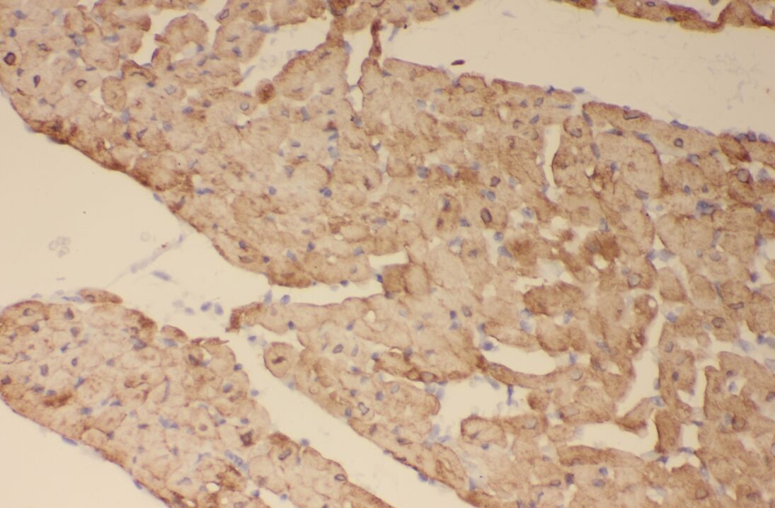 Immunohistochemistry of paraffin-embedded human heart using FNab08100(SOCS3 antibody) at dilution of 1:50
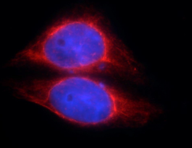 Immunofluorescent analysis of HepG2 cells, using FNab08100(SOCS3 antibody)at 1:10 dilution and Rhodamine-labeled goat anti-rabbit IgG (red). Blue pseudocolor = DAPI (fluorescent DNA dye).
- Background
- SOCS family proteins form part of a classical negative feedback system that regulates cytokine signal transduction. SOCS3 is involved in negative regulation of cytokines that signal through the JAK/STAT pathway. Inhibits cytokine signal transduction by binding to tyrosine kinase receptors including gp130, LIF, erythropoietin, insulin, IL12, GCSF and leptin receptors. Binding to JAK2 inhibits its kinase activity. Suppresses fetal liver erythropoiesis. Regulates onset and maintenance of allergic responses mediated by T-helper type 2 cells. Regulates IL-6 signaling in vivo(By similarity). Probable substrate recognition component of a SCF-like ECS(Elongin BC-CUL2/5-SOCS-box protein) E3 ubiquitin-protein ligase complex which mediates the ubiquitination and subsequent proteasomal degradation of target proteins. Seems to recognize IL6ST(By similarity).