Products
RAF1 antibody
| Size | Price |
|---|---|
| 100µg | Inquiry |
Dispatch Time:
About 3 working days
- Product Name
- RAF1 antibody
- Catalogue No.
- FNab07088
- Size
- 100μg
- Form
- liquid
- Purification
- Immunogen affinity purified
- Purity
- ≥95% as determined by SDS-PAGE
- Clonality
- polyclonal
- Isotype
- IgG
- Storage
- PBS with 0.02% sodium azide and 50% glycerol pH 7.3, -20℃ for 12 months(Avoid repeated freeze / thaw cycles.)
Immunogen
- Immunogen
- v-raf-1 murine leukemia viral oncogene homolog 1
- Alternative Names
- RAF proto-oncogene serine/threonine-protein kinase|Proto-oncogene c-RAF (cRaf)|Raf-1|RAF1|RAF antibody
- UniProt ID
- P04049
- Observed MW
- 76 kDa
Application
- Tested Applications
- ELISA, WB, IHC
- Recommended dilution
- WB: 1:500-1:2000; IHC: 1:20-1:200
Validated Images
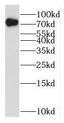 DU 145 cells were subjected to SDS PAGE followed by western blot with FNab07088( RAF1 Antibody) at dilution of 1:1000
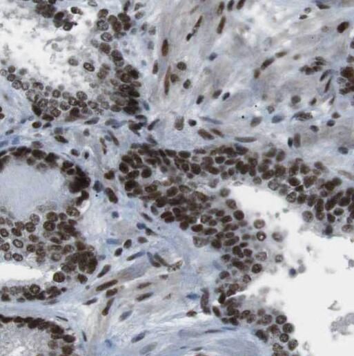 Immunohistochemistry of paraffin-embedded human colon cancer tissue slide using FNab07088(RAF1 Antibody) at dilution of 1:50
- Background
- Serine/threonine-protein kinase that acts as a regulatory link between the membrane-associated Ras GTPases and the MAPK/ERK cascade, and this critical regulatory link functions as a switch determining cell fate decisions including proliferation, differentiation, apoptosis, survival and oncogenic transformation. RAF1 activation initiates a mitogen-activated protein kinase(MAPK) cascade that comprises a sequential phosphorylation of the dual-specific MAPK kinases(MAP2K1/MEK1 and MAP2K2/MEK2) and the extracellular signal-regulated kinases(MAPK3/ERK1 and MAPK1/ERK2). The phosphorylated form of RAF1(on residues Ser-338 and Ser-339, by PAK1) phosphorylates BAD/Bcl2-antagonist of cell death at 'Ser-75'. Phosphorylates adenylyl cyclases: ADCY2, ADCY5 and ADCY6, resulting in their activation. Phosphorylates PPP1R12A resulting in inhibition of the phosphatase activity. Phosphorylates TNNT2/cardiac muscle troponin T. Can promote NF-kB activation and inhibit signal transducers involved in motility(ROCK2), apoptosis(MAP3K5/ASK1 and STK3/MST2), proliferation and angiogenesis(RB1). Can protect cells from apoptosis also by translocating to the mitochondria where it binds BCL2 and displaces BAD/Bcl2-antagonist of cell death. Regulates Rho signaling and migration, and is required for normal wound healing. Plays a role in the oncogenic transformation of epithelial cells via repression of the TJ protein, occludin(OCLN) by inducing the up-regulation of a transcriptional repressor SNAI2/SLUG, which induces down-regulation of OCLN. Restricts caspase activation in response to selected stimuli, notably Fas stimulation, pathogen-mediated macrophage apoptosis, and erythroid differentiation.