Products
PTPRJ antibody
| Size | Price |
|---|---|
| 100µg | Inquiry |
Dispatch Time:
About 3 working days
- Product Name
- PTPRJ antibody
- Catalogue No.
- FNab06945
- Size
- 100μg
- Form
- liquid
- Purification
- Immunogen affinity purified
- Purity
- ≥95% as determined by SDS-PAGE
- Clonality
- polyclonal
- Isotype
- IgG
- Storage
- PBS with 0.02% sodium azide and 50% glycerol pH 7.3, -20℃ for 12 months(Avoid repeated freeze / thaw cycles.)
Immunogen
- Immunogen
- protein tyrosine phosphatase, receptor type, J
- Alternative Names
- Receptor-type tyrosine-protein phosphatase eta (Protein-tyrosine phosphatase eta antibody, R-PTP-eta)|Density-enhanced phosphatase 1 (DEP-1)|HPTP eta|Protein-tyrosine phosphatase receptor type J (R-PTP-J)|PTPRJ|DEP1 antibody
- UniProt ID
- Q12913
- Observed MW
- 146-170 kDa
Application
- Tested Applications
- ELISA, WB
- Recommended dilution
- WB: 1:500-1:2000
Validated Images
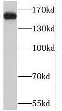 K-562 cells were subjected to SDS PAGE followed by western blot with FNab06945(PTPRJ antibody) at dilution of 1:500
- Background
- Tyrosine phosphatase which dephosphorylates or contributes to the dephosphorylation of CTNND1, FLT3, PDGFRB, MET, RET(variant MEN2A), KDR, LYN, SRC, MAPK1, MAPK3, EGFR, TJP1, OCLN, PIK3R1 and PIK3R2. Plays a role in cell adhesion, migration, proliferation and differentiation. Involved in vascular development. Regulator of macrophage adhesion and spreading. Positively affects cell-matrix adhesion. Positive regulator of platelet activation and thrombosis. Negative regulator of cell proliferation. Negative regulator of PDGF-stimulated cell migration; through dephosphorylation of PDGFR. Positive regulator of endothelial cell survival, as well as of VEGF-induced SRC and AKT activation; through KDR dephosphorylation. Negative regulator of EGFR signaling pathway; through EGFR dephosphorylation. Enhances the barrier function of epithelial junctions during reassembly. Negatively regulates T-cell receptor(TCR) signaling. Upon T-cell TCR activation, it is up-regulated and excluded from the immunological synapses, while upon T-cell-antigen presenting cells(APC) disengagement, it is no longer excluded and can dephosphorylate PLCG1 and LAT to down-regulate prolongation of signaling.