PSMA7 antibody

Synonyms:Proteasome subunit alpha type-7|Proteasome subunit RC6-1|Proteasome subunit XAPC7|PSMA7|HSPC antibody
Catalogue No.:FNab06866Reactivity:Human, Mouse, Rat
Host:RabbitTested Application:ELISA, WB, IHC, FC, IP
Clonality:polyclonalIsotype:IgG
Size Price
100µg Inquiry
Dispatch Time: About 3 working days
  • SPECIFICATIONS
  • FIGURES
  • CONDITIONS
  • FAQS
Product Name
PSMA7 antibody
Catalogue No.
FNab06866
Size
100μg
Form
liquid
Purification
Immunogen affinity purified
Purity
≥95% as determined by SDS-PAGE
Clonality
polyclonal
Isotype
IgG
Storage
PBS with 0.02% sodium azide and 50% glycerol pH 7.3, -20℃ for 12 months(Avoid repeated freeze / thaw cycles.)
Immunogen
Immunogen
proteasome(prosome, macropain) subunit, alpha type, 7
Alternative Names
Proteasome subunit alpha type-7|Proteasome subunit RC6-1|Proteasome subunit XAPC7|PSMA7|HSPC antibody
UniProt ID
O14818
Observed MW
28-30 kDa
Application
Tested Applications
ELISA, WB, IHC, FC, IP
Recommended dilution
WB: 1:500-1:2000; IP: 1:200-1:1000; IHC: 1:20-1:200
Validated Images
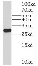
HeLa cells were subjected to SDS PAGE followed by western blot with FNab06866(PSMA7 antibody) at dilution of 1:1000
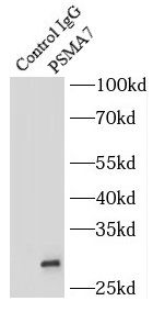
IP Result of anti-PSMA7 (IP:FNab06866, 4ug; Detection:FNab06866 1:500) with mouse brain tissue lysate 4000ug.
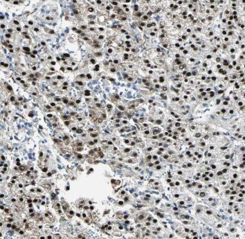
Immunohistochemistry of paraffin-embedded human liver cancer using FNab06866(PSMA7 antibody) at dilution of 1:50
Background
The proteasome is a multicatalytic proteinase complex which is characterized by its ability to cleave peptides with Arg, Phe, Tyr, Leu, and Glu adjacent to the leaving group at neutral or slightly basic pH. The proteasome has an ATP-dependent proteolytic activity. Plays an important role in the regulation of cell proliferation or cell cycle control, transcriptional regulation, immune and stress response, cell differentiation, and apoptosis. Interacts with some important proteins involved in transcription factor regulation, cell cycle transition, viral replication and even tumor initiation and progression. Inhibits the transactivation function of HIF-1A under both normoxic and hypoxia-mimicking conditions. The interaction with EMAP2 increases the proteasome-mediated HIF-1A degradation under the hypoxic conditions. Plays a role in hepatitis C virus internal ribosome entry site-mediated translation. Mediates nuclear translocation of the androgen receptor(AR) and thereby enhances androgen-mediated transactivation. Promotes MAVS degradation and thereby negatively regulates MAVS-mediated innate immune response.
How many times can antibodies be recycled?

First, usually it's not suggested to recycle antibodies. After use, buffer system of antibodies has changed. The storage condition of recycled antibodies for different customers also varies. Thus, the performance efficiency of recycled antibodies can’t be guaranteed. Besides, FineTest ever conducted the antibody recycling assay. Assay results show recycling times of different antibodies also varies. Usually, higher antibody titer allows more repeated use. Customers can determine based on experimental requirements.

Notes: After incubation, we recycle rest antibodies to centrifuge tube and store at 4℃. High titer antibodies can be stored for a minimum of one week. Reuse about three times.

What are components of FineTest antibody buffer?

Components of FineTest antibody buffer are usually PBS with proclin300 or sodium azide, BSA, 50% glycerol. Common preservative is proclin300 or sodium azide, which is widely applied in the lab and industry.

How about the storage temperature and duration of FineTest antibodies?

Most antibodies are stored at -20℃. Directly-labeled flow cytometry antibodies should be stored at 2 - 8℃. The shelf life is one year. If after sales issues for purchased antibodies appear, return or replacement is available. Usually, antibodies can be still used after the one-year warranty. We can offer technical support services.

Is dilution required for FineTest antibodies? What’s the dilute solution?

Directly-labeled flow cytometry antibodies are ready-to-use without dilution. Other antibodies are usually concentrated. Follow the dilution ratio suggested in the manual. Dilute solution for different experiments also varies. Common antibody dilution buffers are acceptable(e.g. PBST, TBST, antibody blocking buffer).

How to retrieve antibodies for immunohistochemistry?

Common retrieval buffers: Tris-EDTA Buffer(pH 9.0); Citrate Buffer(pH 6.0)

Heat induced antibody retrieval:

Method 1: Water-bath heating: Put the beaker with retrieval buffer and slide in the boiling water bath. Keep the boiling state for 15min. Naturally cool to room temperature;

Method 2: Microwave retrieval: Put the beaker with retrieval buffer and slide in the microwave oven. Heat at high power for 5min, Switch OFF for 3min, Heat at medium power for 5min. Naturally cool to room temperature.

How to choose secondary antibodies?

(1) Secondary antibodies react with primary antibodies. Thus, secondary antibodies should be against host species of primary antibodies. E.g. If the primary antibody is derived from rabbit, the relevant secondary antibody should be against rabbit. E.g. goat anti rabbit or donkey anti rabbit.

(2) Choose secondary antibody conjugates according to the experimental type, e.g. ELISA, WB, IHC etc. Common enzyme conjugated secondary antibodies are labelled by HRP, AP etc. Fluorescin or dye labelled secondary antibodies are applied in immunofluorescence and flow cytometry(e.g. FITC, Cy3).

Product Comparison