PIEZO1 antibody

Synonyms:Piezo-type mechanosensitive ion channel component 1|Membrane protein induced by beta-amyloid treatment (Mib)|Protein FAM38A|PIEZO1|FAM38A|KIAA0233 antibody
Catalogue No.:FNab06435Reactivity:Human, Mouse
Host:RabbitTested Application:ELISA, WB,IHC
Clonality:polyclonalIsotype:IgG
Size Price
100µg Inquiry
Dispatch Time: About 3 working days
  • SPECIFICATIONS
  • FIGURES
  • CONDITIONS
  • FAQS
Product Name
PIEZO1 antibody
Catalogue No.
FNab06435
Size
100μg
Form
liquid
Purification
Immunogen affinity purified
Purity
≥95% as determined by SDS-PAGE
Clonality
polyclonal
Isotype
IgG
Storage
PBS with 0.02% sodium azide and 50% glycerol pH 7.3, -20℃ for 12 months(Avoid repeated freeze / thaw cycles.)
Immunogen
Immunogen
family with sequence similarity 38, member A
Alternative Names
Piezo-type mechanosensitive ion channel component 1|Membrane protein induced by beta-amyloid treatment (Mib)|Protein FAM38A|PIEZO1|FAM38A|KIAA0233 antibody
UniProt ID
Q92508
Observed MW
250 kDa
Application
Tested Applications
ELISA, WB,IHC
Recommended dilution
WB: 1:200-1:1000; IHC: 1:50-1:500
Validated Images
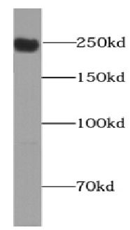
HepG2 cells were subjected to SDS PAGE followed by western blot with FNab06435(Piezo1 antibody) at dilution of 1:500
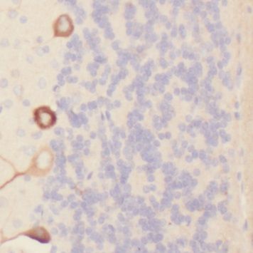
Immunohistochemistry of paraffin-embedded human brain tissue slide using FNab06435(FAM38A Antibody) at dilution of 1:200
Background
Pore-forming subunit of a mechanosensitive non-specific cation channel(PubMed:23479567, PubMed:23695678). Generates currents characterized by a linear current-voltage relationship that are sensitive to ruthenium red and gadolinium. Plays a key role in epithelial cell adhesion by maintaining integrin activation through R-Ras recruitment to the ER, most probably in its activated state, and subsequent stimulation of calpain signaling(PubMed:20016066). In the kidney, may contribute to the detection of intraluminal pressure changes and to urine flow sensing. Acts as shear-stress sensor that promotes endothelial cell organization and alignment in the direction of blood flow through calpain activation(PubMed:25119035). Plays a key role in blood vessel formation and vascular structure in both development and adult physiology(By similarity).
How many times can antibodies be recycled?

First, usually it's not suggested to recycle antibodies. After use, buffer system of antibodies has changed. The storage condition of recycled antibodies for different customers also varies. Thus, the performance efficiency of recycled antibodies can’t be guaranteed. Besides, FineTest ever conducted the antibody recycling assay. Assay results show recycling times of different antibodies also varies. Usually, higher antibody titer allows more repeated use. Customers can determine based on experimental requirements.

Notes: After incubation, we recycle rest antibodies to centrifuge tube and store at 4℃. High titer antibodies can be stored for a minimum of one week. Reuse about three times.

What are components of FineTest antibody buffer?

Components of FineTest antibody buffer are usually PBS with proclin300 or sodium azide, BSA, 50% glycerol. Common preservative is proclin300 or sodium azide, which is widely applied in the lab and industry.

How about the storage temperature and duration of FineTest antibodies?

Most antibodies are stored at -20℃. Directly-labeled flow cytometry antibodies should be stored at 2 - 8℃. The shelf life is one year. If after sales issues for purchased antibodies appear, return or replacement is available. Usually, antibodies can be still used after the one-year warranty. We can offer technical support services.

Is dilution required for FineTest antibodies? What’s the dilute solution?

Directly-labeled flow cytometry antibodies are ready-to-use without dilution. Other antibodies are usually concentrated. Follow the dilution ratio suggested in the manual. Dilute solution for different experiments also varies. Common antibody dilution buffers are acceptable(e.g. PBST, TBST, antibody blocking buffer).

How to retrieve antibodies for immunohistochemistry?

Common retrieval buffers: Tris-EDTA Buffer(pH 9.0); Citrate Buffer(pH 6.0)

Heat induced antibody retrieval:

Method 1: Water-bath heating: Put the beaker with retrieval buffer and slide in the boiling water bath. Keep the boiling state for 15min. Naturally cool to room temperature;

Method 2: Microwave retrieval: Put the beaker with retrieval buffer and slide in the microwave oven. Heat at high power for 5min, Switch OFF for 3min, Heat at medium power for 5min. Naturally cool to room temperature.

How to choose secondary antibodies?

(1) Secondary antibodies react with primary antibodies. Thus, secondary antibodies should be against host species of primary antibodies. E.g. If the primary antibody is derived from rabbit, the relevant secondary antibody should be against rabbit. E.g. goat anti rabbit or donkey anti rabbit.

(2) Choose secondary antibody conjugates according to the experimental type, e.g. ELISA, WB, IHC etc. Common enzyme conjugated secondary antibodies are labelled by HRP, AP etc. Fluorescin or dye labelled secondary antibodies are applied in immunofluorescence and flow cytometry(e.g. FITC, Cy3).

Product Comparison