PEG10 antibody

Synonyms:Retrotransposon-derived protein PEG10|Embryonal carcinoma differentiation-regulated protein|Mammalian retrotransposon-derived protein 2|Myelin expression factor 3-like protein 1 (MEF3-like protein 1)|Paternally expressed gene 10 protein|Retrotransposon gag domain-containing protein 3|Retrotransposon-derived gag-like polyprotein|Ty3/Gypsy-like protein|PEG10|EDR|KIAA1051e|MAR2|MART2|MEF3L1|RGAG3 antibody
Catalogue No.:FNab06302Reactivity:Human, Mouse
Host:RabbitTested Application:ELISA, WB, IHC, IF
Clonality:polyclonalIsotype:IgG
Size Price
100µg Inquiry
Dispatch Time: About 3 working days
  • SPECIFICATIONS
  • FIGURES
  • CONDITIONS
  • FAQS
Product Name
PEG10 antibody
Catalogue No.
FNab06302
Size
100μg
Form
liquid
Purification
Immunogen affinity purified
Purity
≥95% as determined by SDS-PAGE
Clonality
polyclonal
Isotype
IgG
Storage
PBS with 0.02% sodium azide and 50% glycerol pH 7.3, -20℃ for 12 months(Avoid repeated freeze / thaw cycles.)
Immunogen
Immunogen
paternally expressed 10
Alternative Names
Retrotransposon-derived protein PEG10|Embryonal carcinoma differentiation-regulated protein|Mammalian retrotransposon-derived protein 2|Myelin expression factor 3-like protein 1 (MEF3-like protein 1)|Paternally expressed gene 10 protein|Retrotransposon gag domain-containing protein 3|Retrotransposon-derived gag-like polyprotein|Ty3/Gypsy-like protein|PEG10|EDR|KIAA1051e|MAR2|MART2|MEF3L1|RGAG3 antibody
UniProt ID
Q86TG7
Observed MW
50 kDa, 100 kDa
Application
Tested Applications
ELISA, WB, IHC, IF
Recommended dilution
WB: 1:500-1:2000; IHC: 1:20-1:200; IF: 1:20-1:200
Validated Images
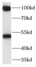
MCF7 cells were subjected to SDS PAGE followed by western blot with FNab06302(PEG10 antibody) at dilution of 1:500
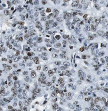
Immunohistochemistry of paraffin-embedded human breast cancer using FNab06302(PEG10 antibody) at dilution of 1:100
Background
Prevents apoptosis in hepatocellular carcinoma(HCC) cells through interaction with SIAH1, a mediator of apoptosis. May also have a role in cell growth promotion and hepatoma formation. Inhibits the TGF-beta signaling by interacting with the TGF-beta receptor ALK1. When overexpressed, induces the formation of cellular extension, such as filipodia in association with ALK1. Involved at the immediate early stage of adipocyte differentiation(By similarity). May bind to the 5'-GCCTGTCTTT-3' DNA sequence of the MB1 domain in the myelin basic protein(MBP) promoter(By similarity).
How many times can antibodies be recycled?

First, usually it's not suggested to recycle antibodies. After use, buffer system of antibodies has changed. The storage condition of recycled antibodies for different customers also varies. Thus, the performance efficiency of recycled antibodies can’t be guaranteed. Besides, FineTest ever conducted the antibody recycling assay. Assay results show recycling times of different antibodies also varies. Usually, higher antibody titer allows more repeated use. Customers can determine based on experimental requirements.

Notes: After incubation, we recycle rest antibodies to centrifuge tube and store at 4℃. High titer antibodies can be stored for a minimum of one week. Reuse about three times.

What are components of FineTest antibody buffer?

Components of FineTest antibody buffer are usually PBS with proclin300 or sodium azide, BSA, 50% glycerol. Common preservative is proclin300 or sodium azide, which is widely applied in the lab and industry.

How about the storage temperature and duration of FineTest antibodies?

Most antibodies are stored at -20℃. Directly-labeled flow cytometry antibodies should be stored at 2 - 8℃. The shelf life is one year. If after sales issues for purchased antibodies appear, return or replacement is available. Usually, antibodies can be still used after the one-year warranty. We can offer technical support services.

Is dilution required for FineTest antibodies? What’s the dilute solution?

Directly-labeled flow cytometry antibodies are ready-to-use without dilution. Other antibodies are usually concentrated. Follow the dilution ratio suggested in the manual. Dilute solution for different experiments also varies. Common antibody dilution buffers are acceptable(e.g. PBST, TBST, antibody blocking buffer).

How to retrieve antibodies for immunohistochemistry?

Common retrieval buffers: Tris-EDTA Buffer(pH 9.0); Citrate Buffer(pH 6.0)

Heat induced antibody retrieval:

Method 1: Water-bath heating: Put the beaker with retrieval buffer and slide in the boiling water bath. Keep the boiling state for 15min. Naturally cool to room temperature;

Method 2: Microwave retrieval: Put the beaker with retrieval buffer and slide in the microwave oven. Heat at high power for 5min, Switch OFF for 3min, Heat at medium power for 5min. Naturally cool to room temperature.

How to choose secondary antibodies?

(1) Secondary antibodies react with primary antibodies. Thus, secondary antibodies should be against host species of primary antibodies. E.g. If the primary antibody is derived from rabbit, the relevant secondary antibody should be against rabbit. E.g. goat anti rabbit or donkey anti rabbit.

(2) Choose secondary antibody conjugates according to the experimental type, e.g. ELISA, WB, IHC etc. Common enzyme conjugated secondary antibodies are labelled by HRP, AP etc. Fluorescin or dye labelled secondary antibodies are applied in immunofluorescence and flow cytometry(e.g. FITC, Cy3).

Product Comparison