Products
NOTCH1 antibody
Research Area:
| Size | Price |
|---|---|
| 100µg | Inquiry |
Dispatch Time:
About 3 working days
- Product Name
- NOTCH1 antibody
- Catalogue No.
- FNab05799
- Size
- 100μg
- Form
- liquid
- Purification
- Immunogen affinity purified
- Purity
- ≥95% as determined by SDS-PAGE
- Clonality
- polyclonal
- Isotype
- IgG
- Storage
- PBS with 0.02% sodium azide and 50% glycerol pH 7.3, -20℃ for 12 months(Avoid repeated freeze / thaw cycles.)
Immunogen
- Immunogen
- Notch homolog 1, translocation-associated(Drosophila)
- Alternative Names
- Neurogenic locus notch homolog protein 1 (Notch 1 antibody, hN1)|Translocation-associated notch protein TAN-1|Notch 1 extracellular truncation (NEXT)|Notch 1 intracellular domain (NICD)|NOTCH1|TAN1 antibody
- UniProt ID
- P46531
- Observed MW
- 270 kDa, 120kd
Application
- Tested Applications
- ELISA, WB, IHC, IP
- Recommended dilution
- WB: 1:500-1:1000; IP: 1:200-1:1000; IHC: 1:50-1:500
Validated Images
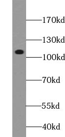 human brain tissue were subjected to SDS PAGE followed by western blot with FNab05799(NOTCH1 antibody) at dilution of 1:500
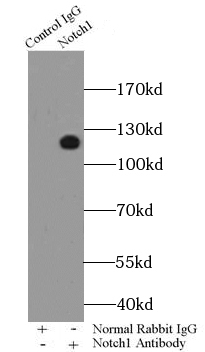 IP Result of anti-Notch1 (IP: FNab05799 4ug; Detection: FNab05799 1:200) with HeLa cells lysate 2000ug.
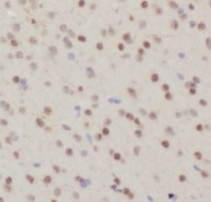 Immunohistochemistry of paraffin-embedded mouse brain tissue tissue slide using FNab05799(NOTCH1 Antibody) at dilution of 1:200
- Background
- Functions as a receptor for membrane-bound ligands Jagged1, Jagged2 and Delta1 to regulate cell-fate determination. Upon ligand activation through the released notch intracellular domain(NICD) it forms a transcriptional activator complex with RBPJ/RBPSUH and activates genes of the enhancer of split locus. Affects the implementation of differentiation, proliferation and apoptotic programs. Involved in angiogenesis; negatively regulates endothelial cell proliferation and migration and angiogenic sprouting. Involved in the maturation of both CD4+ and CD8+ cells in the thymus. Important for follicular differentiation and possibly cell fate selection within the follicle. During cerebellar development, functions as a receptor for neuronal DNER and is involved in the differentiation of Bergmann glia. Represses neuronal and myogenic differentiation. May play an essential role in postimplantation development, probably in some aspect of cell specification and/or differentiation. May be involved in mesoderm development, somite formation and neurogenesis. May enhance HIF1A function by sequestering HIF1AN away from HIF1A. Required for the THBS4 function in regulating protective astrogenesis from the subventricular zone(SVZ) niche after injury. Involved in determination of left/right symmetry by modulating the balance between motile and immotile(sensory) cilia at the left-right organiser(LRO).The antibody is specific to NOTCH1. It can recognize the full length NOTCH1(270 kDa) and all the three cleaved NOTCH1 forms 110-120 kDa.