Products
MSH2 antibody
| Size | Price |
|---|---|
| 100µg | Inquiry |
Dispatch Time:
About 3 working days
- Product Name
- MSH2 antibody
- Catalogue No.
- FNab05372
- Size
- 100μg
- Form
- liquid
- Purification
- Protein A+G purification
- Purity
- ≥95% as determined by SDS-PAGE
- Clonality
- monoclonal
- Isotype
- IgG2a
- Clone ID
- 7C1
- Storage
- PBS with 0.02% sodium azide and 50% glycerol pH 7.3, -20℃ for 12 months(Avoid repeated freeze / thaw cycles.)
Immunogen
- Immunogen
- mutS homolog 2, colon cancer, nonpolyposis type 1
- Alternative Names
- DNA mismatch repair protein Msh2 (hMSH2)|MutS protein homolog 2|MSH2 antibody
- UniProt ID
- P43246
- Observed MW
- 105 kDa
Application
- Tested Applications
- ELISA, WB, IHC
- Recommended dilution
- WB: 1:500-1:2000; IHC: 1:20-1:200
Validated Images
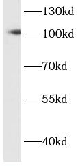 HeLa cells were subjected to SDS PAGE followed by western blot with FNab05372(MSH2 antibody) at dilution of 1:500
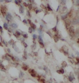 Immunohistochemistry of paraffin-embedded human colon cancer tissue slide using FNab05372( MSH2 Antibody) at dilution of 1:50
- Background
- Component of the post-replicative DNA mismatch repair system(MMR). Forms two different heterodimers: MutS alpha(MSH2-MSH6 heterodimer) and MutS beta(MSH2-MSH3 heterodimer) which binds to DNA mismatches thereby initiating DNA repair. When bound, heterodimers bend the DNA helix and shields approximately 20 base pairs. MutS alpha recognizes single base mismatches and dinucleotide insertion-deletion loops(IDL) in the DNA. MutS beta recognizes larger insertion-deletion loops up to 13 nucleotides long. After mismatch binding, MutS alpha or beta forms a ternary complex with the MutL alpha heterodimer, which is thought to be responsible for directing the downstream MMR events, including strand discrimination, excision, and resynthesis. ATP binding and hydrolysis play a pivotal role in mismatch repair functions. The ATPase activity associated with MutS alpha regulates binding similar to a molecular switch: mismatched DNA provokes ADP-->ATP exchange, resulting in a discernible conformational transition that converts MutS alpha into a sliding clamp capable of hydrolysis-independent diffusion along the DNA backbone. This transition is crucial for mismatch repair. MutS alpha may also play a role in DNA homologous recombination repair. In melanocytes may modulate both UV-B-induced cell cycle regulation and apoptosis.