MAVS antibody

Synonyms:Mitochondrial antiviral-signaling protein (MAVS)|CARD adapter inducing interferon beta (Cardif)|Interferon beta promoter stimulator protein 1 (IPS-1)|Putative NF-kappa-B-activating protein 031N|Virus-induced-signaling adapter (VISA)|MAVS|IPS1|KIAA1271|VISA antibody
Catalogue No.:FNab05032Reactivity:Human, Mouse
Host:RabbitTested Application:ELISA, WB, IF, IP, IHC
Clonality:polyclonalIsotype:IgG
Size Price
100µg Inquiry
Dispatch Time: About 3 working days
  • SPECIFICATIONS
  • FIGURES
  • CONDITIONS
  • FAQS
Product Name
MAVS antibody
Catalogue No.
FNab05032
Size
100μg
Form
liquid
Purification
Immunogen affinity purified
Purity
≥95% as determined by SDS-PAGE
Clonality
polyclonal
Isotype
IgG
Storage
PBS with 0.02% sodium azide and 50% glycerol pH 7.3, -20℃ for 12 months(Avoid repeated freeze / thaw cycles.)
Immunogen
Immunogen
mitochondrial antiviral signaling protein
Alternative Names
Mitochondrial antiviral-signaling protein (MAVS)|CARD adapter inducing interferon beta (Cardif)|Interferon beta promoter stimulator protein 1 (IPS-1)|Putative NF-kappa-B-activating protein 031N|Virus-induced-signaling adapter (VISA)|MAVS|IPS1|KIAA1271|VISA antibody
UniProt ID
Q7Z434
Observed MW
70 kDa
Application
Tested Applications
ELISA, WB, IF, IP, IHC
Recommended dilution
WB: 1:1000-1:4000; IP: 1:500-1:2000; IHC: 1:100-1:500; IF: 1:10-1:100
Validated Images
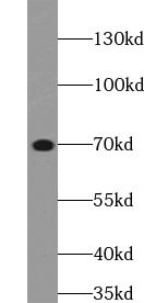
HEK-293 cells were subjected to SDS PAGE followed by western blot with FNab05032(MAVS antibody) at dilution of 1:2000
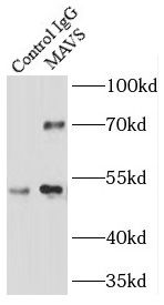
IP Result of anti-MAVS; VISA (IP:FNab05032, 3ug; Detection:FNab05032 1:1000) with HEK-293 cells lysate 1700ug.
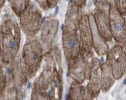
Immunohistochemistry of paraffin-embedded human heart tissue slide using FNab05032(MAVS Antibody) at dilution of 1:100
Background
Required for innate immune defense against viruses. Acts downstream of DDX58/RIG-I and IFIH1/MDA5, which detect intracellular dsRNA produced during viral replication, to coordinate pathways leading to the activation of NF-kappa-B, IRF3 and IRF7, and to the subsequent induction of antiviral cytokines such as IFN-beta and RANTES(CCL5). Peroxisomal and mitochondrial MAVS act sequentially to create an antiviral cellular state. Upon viral infection, peroxisomal MAVS induces the rapid interferon-independent expression of defense factors that provide short-term protection, whereas mitochondrial MAVS activates an interferon-dependent signaling pathway with delayed kinetics, which amplifies and stabilizes the antiviral response. May activate the same pathways following detection of extracellular dsRNA by TLR3. May protect cells from apoptosis.It can undergoe phosphorylation on multiple sites and ubiquitination, which may together cause the molecular weight migrate to about 70 kDa despite the predicated 57 kDa.
How many times can antibodies be recycled?

First, usually it's not suggested to recycle antibodies. After use, buffer system of antibodies has changed. The storage condition of recycled antibodies for different customers also varies. Thus, the performance efficiency of recycled antibodies can’t be guaranteed. Besides, FineTest ever conducted the antibody recycling assay. Assay results show recycling times of different antibodies also varies. Usually, higher antibody titer allows more repeated use. Customers can determine based on experimental requirements.

Notes: After incubation, we recycle rest antibodies to centrifuge tube and store at 4℃. High titer antibodies can be stored for a minimum of one week. Reuse about three times.

What are components of FineTest antibody buffer?

Components of FineTest antibody buffer are usually PBS with proclin300 or sodium azide, BSA, 50% glycerol. Common preservative is proclin300 or sodium azide, which is widely applied in the lab and industry.

How about the storage temperature and duration of FineTest antibodies?

Most antibodies are stored at -20℃. Directly-labeled flow cytometry antibodies should be stored at 2 - 8℃. The shelf life is one year. If after sales issues for purchased antibodies appear, return or replacement is available. Usually, antibodies can be still used after the one-year warranty. We can offer technical support services.

Is dilution required for FineTest antibodies? What’s the dilute solution?

Directly-labeled flow cytometry antibodies are ready-to-use without dilution. Other antibodies are usually concentrated. Follow the dilution ratio suggested in the manual. Dilute solution for different experiments also varies. Common antibody dilution buffers are acceptable(e.g. PBST, TBST, antibody blocking buffer).

How to retrieve antibodies for immunohistochemistry?

Common retrieval buffers: Tris-EDTA Buffer(pH 9.0); Citrate Buffer(pH 6.0)

Heat induced antibody retrieval:

Method 1: Water-bath heating: Put the beaker with retrieval buffer and slide in the boiling water bath. Keep the boiling state for 15min. Naturally cool to room temperature;

Method 2: Microwave retrieval: Put the beaker with retrieval buffer and slide in the microwave oven. Heat at high power for 5min, Switch OFF for 3min, Heat at medium power for 5min. Naturally cool to room temperature.

How to choose secondary antibodies?

(1) Secondary antibodies react with primary antibodies. Thus, secondary antibodies should be against host species of primary antibodies. E.g. If the primary antibody is derived from rabbit, the relevant secondary antibody should be against rabbit. E.g. goat anti rabbit or donkey anti rabbit.

(2) Choose secondary antibody conjugates according to the experimental type, e.g. ELISA, WB, IHC etc. Common enzyme conjugated secondary antibodies are labelled by HRP, AP etc. Fluorescin or dye labelled secondary antibodies are applied in immunofluorescence and flow cytometry(e.g. FITC, Cy3).

Product Comparison