Products
LEO1 antibody
| Size | Price |
|---|---|
| 100µg | Inquiry |
Dispatch Time:
About 3 working days
- Product Name
- LEO1 antibody
- Catalogue No.
- FNab04750
- Size
- 100μg
- Form
- liquid
- Purification
- Immunogen affinity purified
- Purity
- ≥95% as determined by SDS-PAGE
- Clonality
- polyclonal
- Isotype
- IgG
- Storage
- PBS with 0.02% sodium azide and 50% glycerol pH 7.3, -20℃ for 12 months(Avoid repeated freeze / thaw cycles.)
Immunogen
- Immunogen
- Leo1, Paf1/RNA polymerase II complex component, homolog
- Alternative Names
- RNA polymerase-associated protein LEO1|Replicative senescence down-regulated leo1-like protein|LEO1|RDL antibody
- UniProt ID
- Q8WVC0
- Observed MW
- 105 kDa
Application
- Tested Applications
- ELISA, WB, IF, IHC
- Recommended dilution
- WB: 1:500-1:2000; IHC: 1:20-1:200; IF: 1:20-1:200
Validated Images
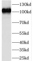 mouse heart tissue were subjected to SDS PAGE followed by western blot with FNab04750(LEO1 antibody) at dilution of 1:500
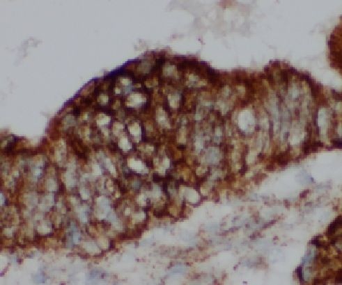 Immunohistochemistry of paraffin-embedded human colon tissue slide using FNab04750(LEO1 Antibody) at dilution of 1:50
- Background
- Component of the PAF1 complex(PAF1C) which has multiple functions during transcription by RNA polymerase II and is implicated in regulation of development and maintenance of embryonic stem cell pluripotency. PAF1C associates with RNA polymerase II through interaction with POLR2A CTD non-phosphorylated and 'Ser-2'-and 'Ser-5'-phosphorylated forms and is involved in transcriptional elongation, acting both indepentently and synergistically with TCEA1 and in cooperation with the DSIF complex and HTATSF1. PAF1C is required for transcription of Hox and Wnt target genes. PAF1C is involved in hematopoiesis and stimulates transcriptional activity of KMT2A/MLL1; it promotes leukemogenesis through association with KMT2A/MLL1-rearranged oncoproteins, such as KMT2A/MLL1-MLLT3/AF9 and KMT2A/MLL1-MLLT1/ENL. PAF1C is involved in histone modifications such as ubiquitination of histone H2B and methylation on histone H3 'Lys-4'(H3K4me3). PAF1C recruits the RNF20/40 E3 ubiquitin-protein ligase complex and the E2 enzyme UBE2A or UBE2B to chromatin which mediate monoubiquitination of 'Lys-120' of histone H2B(H2BK120ub1); UB2A/B-mediated H2B ubiquitination is proposed to be coupled to transcription. PAF1C is involved in mRNA 3' end formation probably through association with cleavage and poly(A) factors. In case of infection by influenza A strain H3N2, PAF1C associates with viral NS1 protein, thereby regulating gene transcription. Involved in polyadenylation of mRNA precursors. Connects PAF1C to Wnt signaling.