ITK antibody

Synonyms:Tyrosine-protein kinase ITK/TSK|Interleukin-2-inducible T-cell kinase (IL-2-inducible T-cell kinase)|Kinase EMT|T-cell-specific kinase|Tyrosine-protein kinase Lyk|ITK|EMT|LYK antibody
Catalogue No.:FNab04414Reactivity:Human, Mouse, Rat
Host:RabbitTested Application:ELISA, WB
Clonality:polyclonalIsotype:IgG
Size Price
100µg Inquiry
Dispatch Time: About 3 working days
  • SPECIFICATIONS
  • FIGURES
  • CONDITIONS
  • FAQS
Product Name
ITK antibody
Catalogue No.
FNab04414
Size
100μg
Form
liquid
Purification
Immunogen affinity purified
Purity
≥95% as determined by SDS-PAGE
Clonality
polyclonal
Isotype
IgG
Storage
PBS with 0.02% sodium azide and 50% glycerol pH 7.3, -20℃ for 12 months(Avoid repeated freeze / thaw cycles.)
Immunogen
Immunogen
IL2-inducible T-cell kinase
Alternative Names
Tyrosine-protein kinase ITK/TSK|Interleukin-2-inducible T-cell kinase (IL-2-inducible T-cell kinase)|Kinase EMT|T-cell-specific kinase|Tyrosine-protein kinase Lyk|ITK|EMT|LYK antibody
UniProt ID
Q08881
Observed MW
68-72 kDa
Application
Tested Applications
ELISA, WB
Recommended dilution
WB: 1:500-1:2000
Validated Images
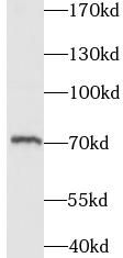
Jurkat cells were subjected to SDS PAGE followed by western blot with FNab04414(ITK antibody) at dilution of 1:1000
Background
Tyrosine kinase that plays an essential role in regulation of the adaptive immune response. Regulates the development, function and differentiation of conventional T-cells and nonconventional NKT-cells. When antigen presenting cells(APC) activate T-cell receptor(TCR), a series of phosphorylation lead to the recruitment of ITK to the cell membrane, in the vicinity of the stimulated TCR receptor, where it is phosphorylated by LCK. Phosphorylation leads to ITK autophosphorylation and full activation. Once activated, phosphorylates PLCG1, leading to the activation of this lipase and subsequent cleavage of its substrates. In turn, the endoplasmic reticulum releases calcium in the cytoplasm and the nuclear activator of activated T-cells(NFAT) translocates into the nucleus to perform its transcriptional duty. Phosphorylates 2 essential adapter proteins: the linker for activation of T-cells/LAT protein and LCP2. Then, a large number of signaling molecules such as VAV1 are recruited and ultimately lead to lymphokine production, T-cell proliferation and differentiation.
How many times can antibodies be recycled?

First, usually it's not suggested to recycle antibodies. After use, buffer system of antibodies has changed. The storage condition of recycled antibodies for different customers also varies. Thus, the performance efficiency of recycled antibodies can’t be guaranteed. Besides, FineTest ever conducted the antibody recycling assay. Assay results show recycling times of different antibodies also varies. Usually, higher antibody titer allows more repeated use. Customers can determine based on experimental requirements.

Notes: After incubation, we recycle rest antibodies to centrifuge tube and store at 4℃. High titer antibodies can be stored for a minimum of one week. Reuse about three times.

What are components of FineTest antibody buffer?

Components of FineTest antibody buffer are usually PBS with proclin300 or sodium azide, BSA, 50% glycerol. Common preservative is proclin300 or sodium azide, which is widely applied in the lab and industry.

How about the storage temperature and duration of FineTest antibodies?

Most antibodies are stored at -20℃. Directly-labeled flow cytometry antibodies should be stored at 2 - 8℃. The shelf life is one year. If after sales issues for purchased antibodies appear, return or replacement is available. Usually, antibodies can be still used after the one-year warranty. We can offer technical support services.

Is dilution required for FineTest antibodies? What’s the dilute solution?

Directly-labeled flow cytometry antibodies are ready-to-use without dilution. Other antibodies are usually concentrated. Follow the dilution ratio suggested in the manual. Dilute solution for different experiments also varies. Common antibody dilution buffers are acceptable(e.g. PBST, TBST, antibody blocking buffer).

How to retrieve antibodies for immunohistochemistry?

Common retrieval buffers: Tris-EDTA Buffer(pH 9.0); Citrate Buffer(pH 6.0)

Heat induced antibody retrieval:

Method 1: Water-bath heating: Put the beaker with retrieval buffer and slide in the boiling water bath. Keep the boiling state for 15min. Naturally cool to room temperature;

Method 2: Microwave retrieval: Put the beaker with retrieval buffer and slide in the microwave oven. Heat at high power for 5min, Switch OFF for 3min, Heat at medium power for 5min. Naturally cool to room temperature.

How to choose secondary antibodies?

(1) Secondary antibodies react with primary antibodies. Thus, secondary antibodies should be against host species of primary antibodies. E.g. If the primary antibody is derived from rabbit, the relevant secondary antibody should be against rabbit. E.g. goat anti rabbit or donkey anti rabbit.

(2) Choose secondary antibody conjugates according to the experimental type, e.g. ELISA, WB, IHC etc. Common enzyme conjugated secondary antibodies are labelled by HRP, AP etc. Fluorescin or dye labelled secondary antibodies are applied in immunofluorescence and flow cytometry(e.g. FITC, Cy3).

Product Comparison