FAS antibody

11th Anniversary Sale
Synonyms:Tumor necrosis factor receptor superfamily member 6|Apo-1 antigen|Apoptosis-mediating surface antigen FAS|FASLG receptor|FAS|APT1|FAS1|TNFRSF6 antibody
Catalogue No.:FNab03016Reactivity:Human, Mouse, Rat
Host:RabbitTested Application:ELISA, WB, IHC
Clonality:polyclonalIsotype:IgG
Size Price
100µg Inquiry
Dispatch Time: About 3 working days
  • SPECIFICATIONS
  • FIGURES
  • CONDITIONS
  • FAQS
Product Name
FAS antibody
Catalogue No.
FNab03016
Size
100μg
Form
liquid
Purification
Immunogen affinity purified
Purity
≥95% as determined by SDS-PAGE
Clonality
polyclonal
Isotype
IgG
Storage
PBS with 0.02% sodium azide and 50% glycerol pH 7.3, -20℃ for 12 months (Avoid repeated freeze / thaw cycles.)
Immunogen
Immunogen
Fas (TNF receptor superfamily, member 6)
Alternative Names
Tumor necrosis factor receptor superfamily member 6|Apo-1 antigen|Apoptosis-mediating surface antigen FAS|FASLG receptor|FAS|APT1|FAS1|TNFRSF6 antibody
UniProt ID
P25445
Observed MW
Refer to figures
Application
Tested Applications
ELISA, WB, IHC
Recommended dilution
WB: 1:500 - 1:2000; IHC: 1:50 - 1:100
Validated Images
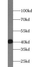
HeLa cells were subjected to SDS PAGE followed by western blot with FNab03016(FAS antibody) at dilution of 1:1000
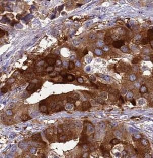
Immunohistochemistry of paraffin-embedded human prostate cancer using FNab03016(FAS antibody) at dilution of 1:100
Background
The protein encoded by this gene is a member of the TNF-receptor superfamily. This receptor contains a death domain. It has been shown to play a central role in the physiological regulation of programmed cell death, and has been implicated in the pathogenesis of various malignancies and diseases of the immune system. The interaction of this receptor with its ligand allows the formation of a death-inducing signaling complex that includes Fas-associated death domain protein (FADD), caspase 8, and caspase 10. The autoproteolytic processing of the caspases in the complex triggers a downstream caspase cascade, and leads to apoptosis. This receptor has been also shown to activate NF-kappaB, MAPK3/ERK1, and MAPK8/JNK, and is found to be involved in transducing the proliferating signals in normal diploid fibroblast and T cells. Several alternatively spliced transcript variants have been described, some of which are candidates for nonsense-mediated mRNA decay (NMD). The isoforms lacking the transmembrane domain may negatively regulate the apoptosis mediated by the full length isoform.
How many times can antibodies be recycled?

First, usually it's not suggested to recycle antibodies. After use, buffer system of antibodies has changed. The storage condition of recycled antibodies for different customers also varies. Thus, the performance efficiency of recycled antibodies can’t be guaranteed. Besides, FineTest ever conducted the antibody recycling assay. Assay results show recycling times of different antibodies also varies. Usually, higher antibody titer allows more repeated use. Customers can determine based on experimental requirements.

Notes: After incubation, we recycle rest antibodies to centrifuge tube and store at 4℃. High titer antibodies can be stored for a minimum of one week. Reuse about three times.

What are components of FineTest antibody buffer?

Components of FineTest antibody buffer are usually PBS with proclin300 or sodium azide, BSA, 50% glycerol. Common preservative is proclin300 or sodium azide, which is widely applied in the lab and industry.

How about the storage temperature and duration of FineTest antibodies?

Most antibodies are stored at -20℃. Directly-labeled flow cytometry antibodies should be stored at 2 - 8℃. The shelf life is one year. If after sales issues for purchased antibodies appear, return or replacement is available. Usually, antibodies can be still used after the one-year warranty. We can offer technical support services.

Is dilution required for FineTest antibodies? What’s the dilute solution?

Directly-labeled flow cytometry antibodies are ready-to-use without dilution. Other antibodies are usually concentrated. Follow the dilution ratio suggested in the manual. Dilute solution for different experiments also varies. Common antibody dilution buffers are acceptable(e.g. PBST, TBST, antibody blocking buffer).

How to retrieve antibodies for immunohistochemistry?

Common retrieval buffers: Tris-EDTA Buffer(pH 9.0); Citrate Buffer(pH 6.0)

Heat induced antibody retrieval:

Method 1: Water-bath heating: Put the beaker with retrieval buffer and slide in the boiling water bath. Keep the boiling state for 15min. Naturally cool to room temperature;

Method 2: Microwave retrieval: Put the beaker with retrieval buffer and slide in the microwave oven. Heat at high power for 5min, Switch OFF for 3min, Heat at medium power for 5min. Naturally cool to room temperature.

How to choose secondary antibodies?

(1) Secondary antibodies react with primary antibodies. Thus, secondary antibodies should be against host species of primary antibodies. E.g. If the primary antibody is derived from rabbit, the relevant secondary antibody should be against rabbit. E.g. goat anti rabbit or donkey anti rabbit.

(2) Choose secondary antibody conjugates according to the experimental type, e.g. ELISA, WB, IHC etc. Common enzyme conjugated secondary antibodies are labelled by HRP, AP etc. Fluorescin or dye labelled secondary antibodies are applied in immunofluorescence and flow cytometry(e.g. FITC, Cy3).

Product Comparison