Products
EIF4A3 antibody
Research Area:
| Size | Price |
|---|---|
| 100µg | Inquiry |
Dispatch Time:
About 3 working days
- Product Name
- EIF4A3 antibody
- Catalogue No.
- FNab02719
- Size
- 100μg
- Form
- liquid
- Purification
- Immunogen affinity purified
- Purity
- ≥95% as determined by SDS-PAGE
- Clonality
- polyclonal
- Isotype
- IgG
- Storage
- PBS with 0.02% sodium azide and 50% glycerol pH 7.3, -20℃ for 12 months(Avoid repeated freeze / thaw cycles.)
Immunogen
- Immunogen
- eukaryotic translation initiation factor 4A, isoform 3
- Alternative Names
- Eukaryotic initiation factor 4A-III (eIF-4A-III antibody, eIF4A-III)|ATP-dependent RNA helicase DDX48|ATP-dependent RNA helicase eIF4A-3|DEAD box protein 48|Eukaryotic initiation factor 4A-like NUK-34|Eukaryotic translation initiation factor 4A isoform 3|Nuclear matrix protein 265 (NMP 265 antibody, hNMP 265)|Eukaryotic initiation factor 4A-III antibody, N-terminally processed|EIF4A3|DDX48|KIAA0111 antibody
- UniProt ID
- P38919
- Observed MW
- 47 kDa
Application
- Tested Applications
- ELISA, WB, IHC, IF, IP
- Recommended dilution
- WB: 1:500-1:2000; IP: 1:200-1:1000; IHC: 1:20-1:200; IF: 1:20-1:200
Validated Images
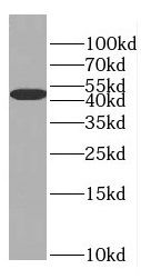 A549 cells were subjected to SDS PAGE followed by western blot with FNab02719(EIF4A3 antibody) at dilution of 1:600
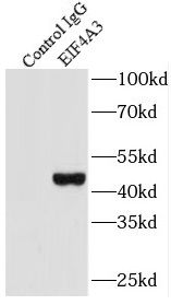 IP Result of anti-EIF4A3 (IP:FNab02719, 3ug; Detection:FNab02719 1:300) with HEK-293 cells lysate 2800ug.
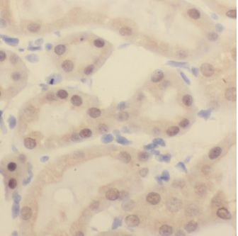 Immunohistochemistry of paraffin-embedded human kidney using FNab02719(EIF4A3 antibody) at dilution of 1:100
- Background
- ATP-dependent RNA helicase. Core component of the splicing-dependent multiprotein exon junction complex(EJC) deposited at splice junctions on mRNAs. The EJC is a dynamic structure consisting of core proteins and several peripheral nuclear and cytoplasmic associated factors that join the complex only transiently either during EJC assembly or during subsequent mRNA metabolism. The EJC marks the position of the exon-exon junction in the mature mRNA for the gene expression machinery and the core components remain bound to spliced mRNAs throughout all stages of mRNA metabolism thereby influencing downstream processes including nuclear mRNA export, subcellular mRNA localization, translation efficiency and nonsense-mediated mRNA decay(NMD). Its RNA-dependent ATPase and RNA-helicase activities are induced by CASC3, but abolished in presence of the MAGOH-RBM8A heterodimer, thereby trapping the ATP-bound EJC core onto spliced mRNA in a stable conformation. The inhibition of ATPase activity by the MAGOH-RBM8A heterodimer increases the RNA-binding affinity of the EJC. Involved in translational enhancement of spliced mRNAs after formation of the 80S ribosome complex. Binds spliced mRNA in sequence-independent manner, 20-24 nucleotides upstream of mRNA exon-exon junctions. Shows higher affinity for single-stranded RNA in an ATP-bound core EJC complex than after the ATP is hydrolyzed. Involved in the splicing modulation of BCL2L1/Bcl-X(and probably other apoptotic genes); specifically inhibits formation of proapoptotic isoforms such as Bcl-X(S); the function is different from the established EJC assembly. Involved in craniofacial development.