DCLRE1C antibody

Synonyms:Protein artemis|DNA cross-link repair 1C protein|Protein A-SCID|SNM1 homolog C (hSNM1C)|SNM1-like protein|DCLRE1C|ARTEMIS|ASCID|SCIDA|SNM1C antibody
Catalogue No.:FNab02268Reactivity:Human, Mouse, Rat
Host:RabbitTested Application:ELISA, WB
Clonality:polyclonalIsotype:IgG
Size Price
100µg Inquiry
Dispatch Time: About 3 working days
  • SPECIFICATIONS
  • FIGURES
  • CONDITIONS
  • FAQS
Product Name
DCLRE1C antibody
Catalogue No.
FNab02268
Size
100μg
Form
liquid
Purification
Immunogen affinity purified
Purity
≥95% as determined by SDS-PAGE
Clonality
polyclonal
Isotype
IgG
Storage
PBS with 0.02% sodium azide and 50% glycerol pH 7.3, -20℃ for 12 months(Avoid repeated freeze / thaw cycles.)
Immunogen
Immunogen
DNA cross-link repair 1C(PSO2 homolog, S. cerevisiae)
Alternative Names
Protein artemis|DNA cross-link repair 1C protein|Protein A-SCID|SNM1 homolog C (hSNM1C)|SNM1-like protein|DCLRE1C|ARTEMIS|ASCID|SCIDA|SNM1C antibody
UniProt ID
Q96SD1
Observed MW
60 kDa
Application
Tested Applications
ELISA, WB
Recommended dilution
WB: 1:500-1:2000
Validated Images
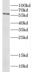
HeLa cells were subjected to SDS PAGE followed by western blot with FNab02268(DCLRE1C antibody) at dilution of 1:400
Background
Required for V(D)J recombination, the process by which exons encoding the antigen-binding domains of immunoglobulins and T-cell receptor proteins are assembled from individual V,(D), and J gene segments. V(D)J recombination is initiated by the lymphoid specific RAG endonuclease complex, which generates site specific DNA double strand breaks(DSBs). These DSBs present two types of DNA end structures: hairpin sealed coding ends and phosphorylated blunt signal ends. These ends are independently repaired by the non homologous end joining(NHEJ) pathway to form coding and signal joints respectively. This protein exhibits single-strand specific 5'-3' exonuclease activity in isolation and acquires endonucleolytic activity on 5' and 3' hairpins and overhangs when in a complex with PRKDC. The latter activity is required specifically for the resolution of closed hairpins prior to the formation of the coding joint. May also be required for the repair of complex DSBs induced by ionizing radiation, which require substantial end-processing prior to religation by NHEJ.
How many times can antibodies be recycled?

First, usually it's not suggested to recycle antibodies. After use, buffer system of antibodies has changed. The storage condition of recycled antibodies for different customers also varies. Thus, the performance efficiency of recycled antibodies can’t be guaranteed. Besides, FineTest ever conducted the antibody recycling assay. Assay results show recycling times of different antibodies also varies. Usually, higher antibody titer allows more repeated use. Customers can determine based on experimental requirements.

Notes: After incubation, we recycle rest antibodies to centrifuge tube and store at 4℃. High titer antibodies can be stored for a minimum of one week. Reuse about three times.

What are components of FineTest antibody buffer?

Components of FineTest antibody buffer are usually PBS with proclin300 or sodium azide, BSA, 50% glycerol. Common preservative is proclin300 or sodium azide, which is widely applied in the lab and industry.

How about the storage temperature and duration of FineTest antibodies?

Most antibodies are stored at -20℃. Directly-labeled flow cytometry antibodies should be stored at 2 - 8℃. The shelf life is one year. If after sales issues for purchased antibodies appear, return or replacement is available. Usually, antibodies can be still used after the one-year warranty. We can offer technical support services.

Is dilution required for FineTest antibodies? What’s the dilute solution?

Directly-labeled flow cytometry antibodies are ready-to-use without dilution. Other antibodies are usually concentrated. Follow the dilution ratio suggested in the manual. Dilute solution for different experiments also varies. Common antibody dilution buffers are acceptable(e.g. PBST, TBST, antibody blocking buffer).

How to retrieve antibodies for immunohistochemistry?

Common retrieval buffers: Tris-EDTA Buffer(pH 9.0); Citrate Buffer(pH 6.0)

Heat induced antibody retrieval:

Method 1: Water-bath heating: Put the beaker with retrieval buffer and slide in the boiling water bath. Keep the boiling state for 15min. Naturally cool to room temperature;

Method 2: Microwave retrieval: Put the beaker with retrieval buffer and slide in the microwave oven. Heat at high power for 5min, Switch OFF for 3min, Heat at medium power for 5min. Naturally cool to room temperature.

How to choose secondary antibodies?

(1) Secondary antibodies react with primary antibodies. Thus, secondary antibodies should be against host species of primary antibodies. E.g. If the primary antibody is derived from rabbit, the relevant secondary antibody should be against rabbit. E.g. goat anti rabbit or donkey anti rabbit.

(2) Choose secondary antibody conjugates according to the experimental type, e.g. ELISA, WB, IHC etc. Common enzyme conjugated secondary antibodies are labelled by HRP, AP etc. Fluorescin or dye labelled secondary antibodies are applied in immunofluorescence and flow cytometry(e.g. FITC, Cy3).

Product Comparison