Products
CSNK2A1 antibody
| Size | Price |
|---|---|
| 100µg | Inquiry |
Dispatch Time:
About 3 working days
- Product Name
- CSNK2A1 antibody
- Catalogue No.
- FNab02023
- Size
- 100μg
- Form
- liquid
- Purification
- Immunogen affinity purified
- Purity
- ≥95% as determined by SDS-PAGE
- Clonality
- polyclonal
- Isotype
- IgG
- Storage
- PBS with 0.02% sodium azide and 50% glycerol pH 7.3, -20℃ for 12 months(Avoid repeated freeze / thaw cycles.)
Immunogen
- Immunogen
- casein kinase 2, alpha 1 polypeptide
- Alternative Names
- Casein kinase II subunit alpha (CK II alpha)|CSNK2A1|CK2A1 antibody
- UniProt ID
- P68400
- Observed MW
- 45 kDa
Application
- Tested Applications
- ELISA, WB, IHC, IF, IP
- Recommended dilution
- WB: 1:500-1:2000; IP: 1:500-1:2000; IHC: 1:20-1:200; IF: 1:10-1:100
Validated Images
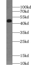 HeLa cells were subjected to SDS PAGE followed by western blot with FNab02023(CSNK2A1 antibody) at dilution of 1:1000
 IP Result of anti-CSNK2A1 (IP:FNab02023, 4ug; Detection:FNab02023 1:1000) with HeLa cells lysate 2800ug.
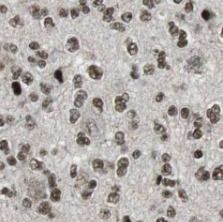 Immunohistochemistry of paraffin-embedded human gliomas using FNab02023(CSNK2A1 antibody) at dilution of 1:100
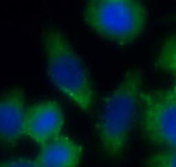 Immunofluorescent analysis of HeLa cells using FNab02023 (CSNK2A1 antibody) at dilution of 1:25 and Alexa Fluor 488-conjugated Goat Anti-Rabbit IgG(H+L)
- Background
- Catalytic subunit of a constitutively active serine/threonine-protein kinase complex that phosphorylates a large number of substrates containing acidic residues C-terminal to the phosphorylated serine or threonine. Regulates numerous cellular processes, such as cell cycle progression, apoptosis and transcription, as well as viral infection. May act as a regulatory node which integrates and coordinates numerous signals leading to an appropriate cellular response. During mitosis, functions as a component of the p53/TP53-dependent spindle assembly checkpoint(SAC) that maintains cyclin-B-CDK1 activity and G2 arrest in response to spindle damage. Also required for p53/TP53-mediated apoptosis, phosphorylating 'Ser-392' of p53/TP53 following UV irradiation. Can also negatively regulate apoptosis. Phosphorylates the caspases CASP9 and CASP2 and the apoptotic regulator NOL3. Phosphorylation protects CASP9 from cleavage and activation by CASP8, and inhibits the dimerization of CASP2 and activation of CASP8. Regulates transcription by direct phosphorylation of RNA polymerases I, II, III and IV. Also phosphorylates and regulates numerous transcription factors including NF-kappa-B, STAT1, CREB1, IRF1, IRF2, ATF1, SRF, MAX, JUN, FOS, MYC and MYB. Phosphorylates Hsp90 and its co-chaperones FKBP4 and CDC37, which is essential for chaperone function. Regulates Wnt signaling by phosphorylating CTNNB1 and the transcription factor LEF1. Acts as an ectokinase that phosphorylates several extracellular proteins. During viral infection, phosphorylates various proteins involved in the viral life cycles of EBV, HSV, HBV, HCV, HIV, CMV and HPV. Phosphorylates PML at 'Ser-565' and primes it for ubiquitin-mediated degradation. Plays an important role in the circadian clock function by phosphorylating ARNTL/BMAL1 at 'Ser-90' which is pivotal for its interaction with CLOCK and which controls CLOCK nuclear entry(PubMed:11239457, PubMed:11704824, PubMed:16193064, PubMed:19188443, PubMed:20625391, PubMed:22406621). Phosphorylates CCAR2 at 'Thr-454' in gastric carcinoma tissue(PubMed:24962073).