COX6A1 antibody

11th Anniversary Sale
Synonyms:Cytochrome c oxidase subunit 6A1 antibody, mitochondrial|Cytochrome c oxidase polypeptide VIa-liver|Cytochrome c oxidase subunit VIA-liver (COX VIa-L)|COX6A1|COX6AL antibody
Catalogue No.:FNab01902Reactivity:Human, Mouse, Rat
Host:RabbitTested Application:ELISA, WB, IHC
Clonality:polyclonalIsotype:IgG
Size Price
100µg Inquiry
Dispatch Time: About 3 working days
  • SPECIFICATIONS
  • FIGURES
  • CONDITIONS
  • FAQS
Product Name
COX6A1 antibody
Catalogue No.
FNab01902
Size
100μg
Form
liquid
Purification
Immunogen affinity purified
Purity
≥95% as determined by SDS-PAGE
Clonality
polyclonal
Isotype
IgG
Storage
PBS with 0.02% sodium azide and 50% glycerol pH 7.3, -20℃ for 12 months (Avoid repeated freeze / thaw cycles.)
Immunogen
Immunogen
cytochrome c oxidase subunit VIa polypeptide 1
Alternative Names
Cytochrome c oxidase subunit 6A1 antibody, mitochondrial|Cytochrome c oxidase polypeptide VIa-liver|Cytochrome c oxidase subunit VIA-liver (COX VIa-L)|COX6A1|COX6AL antibody
UniProt ID
P12074
Observed MW
12 kDa
Application
Tested Applications
ELISA, WB, IHC
Recommended dilution
WB: 1:200 - 1:2000; IHC: 1:20 - 1:200
Validated Images
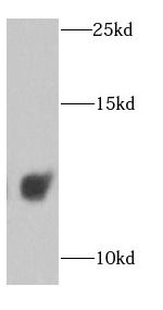
human brain tissue were subjected to SDS PAGE followed by western blot with FNab01902(COX6A1 antibody) at dilution of 1:1000
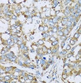
Immunohistochemistry of paraffin-embedded human liver cancer using FNab01902(COX6A1 antibody) at dilution of 1:100
Background
Cytochrome c oxidase (COX), the terminal enzyme of the mitochondrial respiratory chain, catalyzes the electron transfer from reduced cytochrome c to oxygen. It is a heteromeric complex consisting of 3 catalytic subunits encoded by mitochondrial genes and multiple structural subunits encoded by nuclear genes. The mitochondrially-encoded subunits function in the electron transfer and the nuclear-encoded subunits may function in the regulation and assembly of the complex. This nuclear gene encodes polypeptide 1 (liver isoform) of subunit VIa, and polypeptide 1 is found in all non-muscle tissues. Polypeptide 2 (heart/muscle isoform) of subunit VIa is encoded by a different gene, and is present only in striated muscles. These two polypeptides share 66% amino acid sequence identity. It has been reported that there may be several pseudogenes on chromosomes 1, 6, 7q21, 7q31-32 and 12. However, only one pseudogene (COX6A1P) on chromosome 1p31.1 has been documented.
How many times can antibodies be recycled?

First, usually it's not suggested to recycle antibodies. After use, buffer system of antibodies has changed. The storage condition of recycled antibodies for different customers also varies. Thus, the performance efficiency of recycled antibodies can’t be guaranteed. Besides, FineTest ever conducted the antibody recycling assay. Assay results show recycling times of different antibodies also varies. Usually, higher antibody titer allows more repeated use. Customers can determine based on experimental requirements.

Notes: After incubation, we recycle rest antibodies to centrifuge tube and store at 4℃. High titer antibodies can be stored for a minimum of one week. Reuse about three times.

What are components of FineTest antibody buffer?

Components of FineTest antibody buffer are usually PBS with proclin300 or sodium azide, BSA, 50% glycerol. Common preservative is proclin300 or sodium azide, which is widely applied in the lab and industry.

How about the storage temperature and duration of FineTest antibodies?

Most antibodies are stored at -20℃. Directly-labeled flow cytometry antibodies should be stored at 2 - 8℃. The shelf life is one year. If after sales issues for purchased antibodies appear, return or replacement is available. Usually, antibodies can be still used after the one-year warranty. We can offer technical support services.

Is dilution required for FineTest antibodies? What’s the dilute solution?

Directly-labeled flow cytometry antibodies are ready-to-use without dilution. Other antibodies are usually concentrated. Follow the dilution ratio suggested in the manual. Dilute solution for different experiments also varies. Common antibody dilution buffers are acceptable(e.g. PBST, TBST, antibody blocking buffer).

How to retrieve antibodies for immunohistochemistry?

Common retrieval buffers: Tris-EDTA Buffer(pH 9.0); Citrate Buffer(pH 6.0)

Heat induced antibody retrieval:

Method 1: Water-bath heating: Put the beaker with retrieval buffer and slide in the boiling water bath. Keep the boiling state for 15min. Naturally cool to room temperature;

Method 2: Microwave retrieval: Put the beaker with retrieval buffer and slide in the microwave oven. Heat at high power for 5min, Switch OFF for 3min, Heat at medium power for 5min. Naturally cool to room temperature.

How to choose secondary antibodies?

(1) Secondary antibodies react with primary antibodies. Thus, secondary antibodies should be against host species of primary antibodies. E.g. If the primary antibody is derived from rabbit, the relevant secondary antibody should be against rabbit. E.g. goat anti rabbit or donkey anti rabbit.

(2) Choose secondary antibody conjugates according to the experimental type, e.g. ELISA, WB, IHC etc. Common enzyme conjugated secondary antibodies are labelled by HRP, AP etc. Fluorescin or dye labelled secondary antibodies are applied in immunofluorescence and flow cytometry(e.g. FITC, Cy3).

Product Comparison