Products
C1QBP antibody
Research Area:
| Size | Price |
|---|---|
| 100µg | Inquiry |
Dispatch Time:
About 3 working days
- Product Name
- C1QBP antibody
- Catalogue No.
- FNab01073
- Size
- 100μg
- Form
- liquid
- Purification
- Immunogen affinity purified
- Purity
- ≥95% as determined by SDS-PAGE
- Clonality
- polyclonal
- Isotype
- IgG
- Storage
- PBS with 0.02% sodium azide and 50% glycerol pH 7.3, -20℃ for 12 months(Avoid repeated freeze / thaw cycles.)
Immunogen
- Immunogen
- complement component 1, q subcomponent binding protein
- Alternative Names
- Complement component 1 Q subcomponent-binding protein antibody, mitochondrial|ASF/SF2-associated protein p32|Glycoprotein gC1qBP (C1qBP)|Hyaluronan-binding protein 1|Mitochondrial matrix protein p32|gC1q-R protein|p33 (SF2AP32)|C1QBP|GC1QBP|HABP1|SF2P32 antibody
- UniProt ID
- Q07021
- Observed MW
- 32 kDa
Application
- Tested Applications
- ELISA, WB, IHC, IF
- Recommended dilution
- WB: 1:500-1:2000; IHC: 1:20-1:200; IF: 1:20-1:200
Validated Images
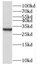 K-562 cells were subjected to SDS PAGE followed by western blot with FNab01073(C1QBP Antibody) at dilution of 1:1000
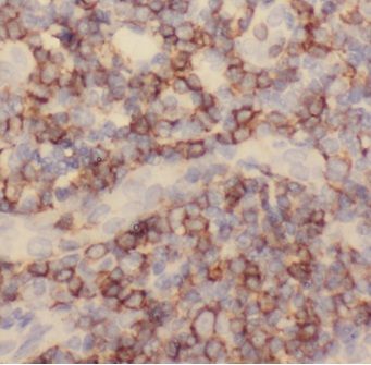 Immunohistochemistry of paraffin-embedded human tonsillitis slide using FNab01073(C1QBP Antibody) at dilution of 1:50
- Background
- Is believed to be a multifunctional and multicompartmental protein involved in inflammation and infection processes, ribosome biogenesis, regulation of apoptosis, transcriptional regulation and pre-mRNA splicing. At the cell surface is thought to act as an endothelial receptor for plasma proteins of the complement and kallikrein-kinin cascades. Putative receptor for C1q; specifically binds to the globular "heads" of C1q thus inhibiting C1; may perform the receptor function through a complex with C1qR/CD93. In complex with cytokeratin-1/KRT1 is a high affinity receptor for kininogen-1/HMWK. Can also bind other plasma proteins, such as coagulation factor XII leading to its autoactivation. May function to bind initially fluid kininogen-1 to the cell membrane. The secreted form may enhance both extrinsic and intrinsic coagulation pathways. It is postulated that the cell surface form requires docking with transmembrane proteins for downstream signaling which might be specific for a cell-type or response. By acting as C1q receptor is involved in chemotaxis of immature dendritic cells and neutrophils and is proposed to signal through CD209/DC-SIGN on immature dendritic cells, through integrin alpha-4/beta-1 during trophoblast invasion of the decidua, and through integrin beta-1 during endothelial cell adhesion and spreading. Signaling involved in inhibition of innate immune response is implicating the PI3K-AKT/PKB pathway. In mitochondrial translation may be involved in formation of functional 55S mitoribosomes; the function seems to involve its RNA-binding activity. May be involved in the nucleolar ribosome maturation process; the function may involve the exchange of FBL for RRP1 in the association with pre-ribosome particles. Involved in regulation of RNA splicing by inhibiting the RNA-binding capacity of SRSF1 and its phosphorylation. Is required for the nuclear translocation of splicing factor U2AF1L4. Involved in regulation of CDKN2A-and HRK-mediated apoptosis. Stabilizes mitochondrial CDKN2A isoform smARF. May be involved in regulation of FOXC1 transcriptional activity and NFY/CCAAT-binding factor complex-mediated transcription. In infection processes acts as an attachment site for microbial proteins, including Listeria monocytogenes internalin B and Staphylococcus aureus protein A. May play a role in antibacterial defense as it can bind to cell surface hyaluronan and inhibit Streptococcus pneumoniae hyaluronate lyase. Involved in replication of Rubella virus. May be involved in modulation of the immune response; ligation by HCV core protein is resulting in suppresion of interleukin-12 production in monocyte-derived dendritic cells. Involved in regulation of antiviral response by inhibiting DDX58-and IFIH1-mediated signaling pathways probably involving its association with MAVS after viral infection. Involved in HIV-1 replication, presumably by contributing to splicing of viral RNA.