ATP5F1A antibody

Synonyms:ATP synthase subunit alpha antibody, mitochondrial|ATP synthase F1 subunit alpha|ATP5F1A|ATP5A|ATP5A1|ATP5AL2|ATPM antibody
Catalogue No.:FNab00703Reactivity:Human, Mouse, Rat
Host:MouseTested Application:ELISA, IHC, IF, WB, FC
Clonality:monoclonalIsotype:IgG1
Size Price
100µg Inquiry
Dispatch Time: About 3 working days
  • SPECIFICATIONS
  • FIGURES
  • CONDITIONS
  • FAQS
Product Name
ATP5F1A antibody
Catalogue No.
FNab00703
Size
100μg
Form
liquid
Purification
Protein A+G purification
Purity
≥95% as determined by SDS-PAGE
Clonality
monoclonal
Isotype
IgG1
Clone ID
9G0
Storage
PBS with 0.02% sodium azide and 50% glycerol pH 7.3, -20℃ for 12 months(Avoid repeated freeze / thaw cycles.)
Immunogen
Immunogen
ATP synthase, H+ transporting, mitochondrial F1 complex, alpha subunit 1, cardiac muscle
Alternative Names
ATP synthase subunit alpha antibody, mitochondrial|ATP synthase F1 subunit alpha|ATP5F1A|ATP5A|ATP5A1|ATP5AL2|ATPM antibody
UniProt ID
P25705
Observed MW
50-55 kDa
Application
Tested Applications
ELISA, IHC, IF, WB, FC
Recommended dilution
WB: 1:500-1:2000; IP: 1:500-1:1000; IHC: 1:20-1:200; IF: 1:20-1:200
Validated Images
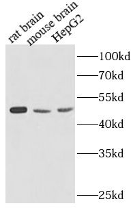
Various lysates were subjected to SDS PAGE followed by western blot with FNab00703(ATP5A1 antibody) at dilution of 1:1000
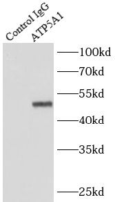
IP Result of anti-ATP5A1 (IP: FNab00703, 5ug; Detection: FNab00703 1:500) with mouse heart tissue lysate 4000ug.
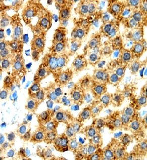
Immunohistochemistry of paraffin-embedded human liver using FNab00703(ATP5A1 antibody) at dilution of 1:50
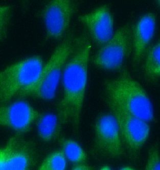
Immunofluorescent analysis of Hela cells using FNab00703 (ATP5A1 antibody) at dilution of 1:25
Background
Mitochondrial membrane ATP synthase(F(1)F(0) ATP synthase or Complex V) produces ATP from ADP in the presence of a proton gradient across the membrane which is generated by electron transport complexes of the respiratory chain. F-type ATPases consist of two structural domains, F(1)-containing the extramembraneous catalytic core, and F(0)-containing the membrane proton channel, linked together by a central stalk and a peripheral stalk. During catalysis, ATP synthesis in the catalytic domain of F(1) is coupled via a rotary mechanism of the central stalk subunits to proton translocation. Subunits alpha and beta form the catalytic core in F(1). Rotation of the central stalk against the surrounding alpha(3)beta(3) subunits leads to hydrolysis of ATP in three separate catalytic sites on the beta subunits. Subunit alpha does not bear the catalytic high-affinity ATP-binding sites(By similarity).
How many times can antibodies be recycled?

First, usually it's not suggested to recycle antibodies. After use, buffer system of antibodies has changed. The storage condition of recycled antibodies for different customers also varies. Thus, the performance efficiency of recycled antibodies can’t be guaranteed. Besides, FineTest ever conducted the antibody recycling assay. Assay results show recycling times of different antibodies also varies. Usually, higher antibody titer allows more repeated use. Customers can determine based on experimental requirements.

Notes: After incubation, we recycle rest antibodies to centrifuge tube and store at 4℃. High titer antibodies can be stored for a minimum of one week. Reuse about three times.

What are components of FineTest antibody buffer?

Components of FineTest antibody buffer are usually PBS with proclin300 or sodium azide, BSA, 50% glycerol. Common preservative is proclin300 or sodium azide, which is widely applied in the lab and industry.

How about the storage temperature and duration of FineTest antibodies?

Most antibodies are stored at -20℃. Directly-labeled flow cytometry antibodies should be stored at 2 - 8℃. The shelf life is one year. If after sales issues for purchased antibodies appear, return or replacement is available. Usually, antibodies can be still used after the one-year warranty. We can offer technical support services.

Is dilution required for FineTest antibodies? What’s the dilute solution?

Directly-labeled flow cytometry antibodies are ready-to-use without dilution. Other antibodies are usually concentrated. Follow the dilution ratio suggested in the manual. Dilute solution for different experiments also varies. Common antibody dilution buffers are acceptable(e.g. PBST, TBST, antibody blocking buffer).

How to retrieve antibodies for immunohistochemistry?

Common retrieval buffers: Tris-EDTA Buffer(pH 9.0); Citrate Buffer(pH 6.0)

Heat induced antibody retrieval:

Method 1: Water-bath heating: Put the beaker with retrieval buffer and slide in the boiling water bath. Keep the boiling state for 15min. Naturally cool to room temperature;

Method 2: Microwave retrieval: Put the beaker with retrieval buffer and slide in the microwave oven. Heat at high power for 5min, Switch OFF for 3min, Heat at medium power for 5min. Naturally cool to room temperature.

How to choose secondary antibodies?

(1) Secondary antibodies react with primary antibodies. Thus, secondary antibodies should be against host species of primary antibodies. E.g. If the primary antibody is derived from rabbit, the relevant secondary antibody should be against rabbit. E.g. goat anti rabbit or donkey anti rabbit.

(2) Choose secondary antibody conjugates according to the experimental type, e.g. ELISA, WB, IHC etc. Common enzyme conjugated secondary antibodies are labelled by HRP, AP etc. Fluorescin or dye labelled secondary antibodies are applied in immunofluorescence and flow cytometry(e.g. FITC, Cy3).

Product Comparison