Products
SH2B1 antibody
| Size | Price |
|---|---|
| 100µg | Inquiry |
Dispatch Time:
About 3 working days
- Product Name
- SH2B1 antibody
- Catalogue No.
- FNab07818
- Size
- 100μg
- Form
- liquid
- Purification
- Immunogen affinity purified
- Purity
- ≥95% as determined by SDS-PAGE
- Clonality
- polyclonal
- Isotype
- IgG
- Storage
- PBS with 0.02% sodium azide and 50% glycerol pH 7.3, -20℃ for 12 months(Avoid repeated freeze / thaw cycles.)
Immunogen
- Immunogen
- SH2B adaptor protein 1
- Alternative Names
- SH2B adapter protein 1|Pro-rich antibody, PH and SH2 domain-containing signaling mediator (PSM)|SH2 domain-containing protein 1B|SH2B1|KIAA1299|SH2B antibody
- UniProt ID
- Q9NRF2
- Observed MW
- 79 kDa
Application
- Tested Applications
- ELISA, WB, IP, IHC, IF
- Recommended dilution
- WB: 1:200-1:2000; IP: 1:200-1:2000; IHC: 1:20-1:200; IF: 1:20-1:200
Validated Images
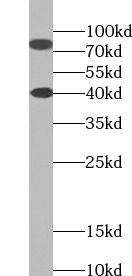 HEK-293 cells were subjected to SDS PAGE followed by western blot with FNab07818(SH2B1 Antibody) at dilution of 1:600
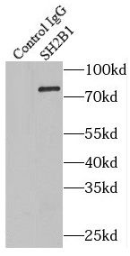 IP Result of anti-SH2B1 (IP:FNab07818, 4ug; Detection:FNab07818 1:600) with HEK-293 cells lysate 3000ug.
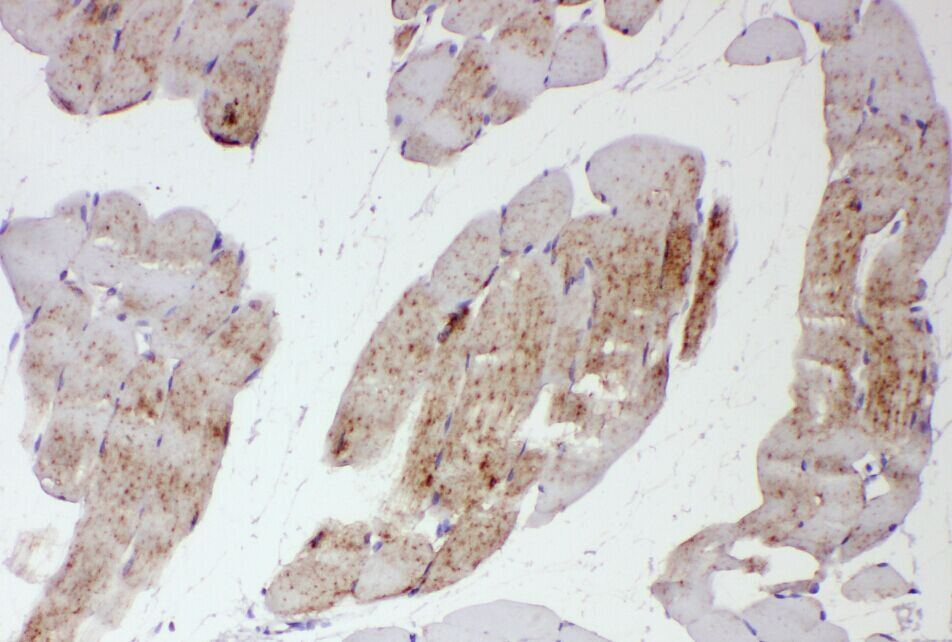 Immunohistochemistry of paraffin-embedded human skeletal muscle tissue slide using FNab07818(SH2B1 Antibody) at dilution of 1:50
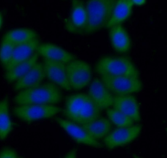 Immunofluorescent analysis of HeLa cells using FNab07818( SH2B1 Antibody) at dilution of 1:50 and Alexa Fluor 488-conjugated AffiniPure Goat Anti-Rabbit IgG(H+L)
- Background
- Adapter protein for several members of the tyrosine kinase receptor family. Involved in multiple signaling pathways mediated by Janus kinase(JAK) and receptor tyrosine kinases, including the receptors of insulin(INS), insulin-like growth factor I(IGF1), nerve growth factor(NGF), brain-derived neurotrophic factor(BDNF), glial cell line-derived neurotrophic factor(GDNF), platelet-derived growth factor(PDGF) and fibroblast growth factors(FGFs). In growth hormone(GH) signaling, autophosphorylated('Tyr-813') JAK2 recruits SH2B1, which in turn is phosphorylated by JAK2 on tyrosine residues. These phosphotyrosines form potential binding sites for other signaling proteins. GH also promotes serine/threonine phosphorylation of SH2B1 and these phosphorylated residues may serve to recruit other proteins to the GHR-JAK2-SH2B1 complexes, such as RAC1. In leptin(LEP) signaling, binds to and potentiates the activation of JAK2 by globally enhancing downstream pathways. In response to leptin, binds simultaneously to both, JAK2 and IRS1 or IRS2, thus mediating formation of a complex of JAK2, SH2B1 and IRS1 or IRS2. Mediates tyrosine phosphorylation of IRS1 and IRS2, resulting in activation of the PI 3-kinase pathway. Acts as positive regulator of NGF-mediated activation of the Akt/Forkhead pathway; prolongs NGF-induced phosphorylation of AKT1 on 'Ser-473' and AKT1 enzymatic activity. Enhances the kinase activity of the cytokine receptor-associated tyrosine kinase JAK2 and of other receptor tyrosine kinases, such as FGFR3 and NTRK1. For JAK2, the mechanism seems to involve dimerization of both, SH2B1 and JAK2. Enhances RET phosphorylation and kinase activity. Isoforms seem to be differentially involved in IGF-I and PDGF-induced mitogenesis(By similarity).