Abstract: Th17 cells are the important subgroup of CD4+ T cells. Pro-inflammatory effects play a key role in autoimmune diseases and inflammatory response. Unlike Treg cell for regulating immune response, Th17 cells mediate immune activation via secreting cytokines(e.g. IL-17). Th17 markers flow cytometry can significantly detect proportion and function of Th17 cells.
Keywords: Th17 Markers, Flow Cytometry, Autoimmune Diseases, Immune Response
1. Components and Functions
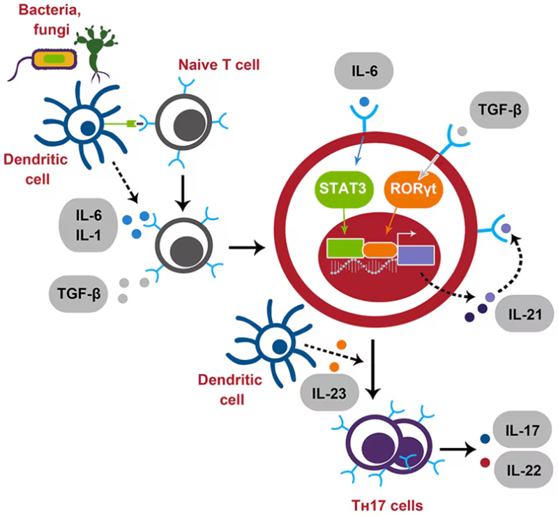
Pro-inflammatory roles of Th17 cells in autoimmune diseases and defence against pathogens are very important. Like CD4+ T cell subgroup(e.g. Th1, Th2), secretion of cytokines activates macrophages, B cells and CD8+ T cell, also requiring involvement of Th17 cells in immune response. The plasticity of Th17 cells allows the conversion to Treg cell under specific conditions. Dynamic equilibrium between them is maintained. Imbalance may cause dysimmunity. The differentiation of Th17 is primarily induced by IL-6, IL-21, IL-23 and TGF-β. Secreted cytokines(e.g. IL-17, IL-21, IL-22 and TNF-α etc) are important in innate immunity and inflammatory response.
2. Common Th17 Markers in Flow Cytometry
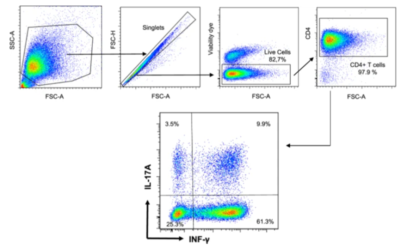
Detection of cytokine IL-17A in mouse sample can recognize Th17 cells. Phenotypic analysis and function evaluation for Th17 cells are an integration of IL-17A, nuclear transcriptional factors(RORγt), surface markers CCR4(CD194) and CCR6(CD196).
Single cell suspension sample of Th17 cells can be obtained from tissues(e.g. periph-eral blood, spleen or lymph node etc). The amount of Th1, Th2 and Th17 in normal physiological conditions are less. Secreted cytokines are relatively lower. Thus, detection of these factors requires for in vitro stimulation. Common stimulators include PMA and Ionomycin. They can activate protein kinase C and induce phosphorylation cascade to further promote activation of T cell and expression of cytokines.
After stimulation, extracellular secretion of cytokines are rapid. Flow cytometry detection of these cytokines uses blocker to reserve in cells. Brefeldin A(BFA) is commonly used to inhibit the transport and secretion of proteins via Golgi apparatus. Retention of cytokines in cells facilitates intracellular staining and flow cytometry analysis.
FineTest also offers stimulation blocking kit(K083), which is widely approved by many customers.
3. Notes
● Blood collection should use heparin sodium anticoagulant to avoid inhibition of EDTA/sodium citrate for cytokine detection.
● PMA stimulation can induce endocytosis of CD4 on T cell surface, and affect detection of CD4+ cell. CD4 can be detected after permeabilization.
● Duration of PMA stimulation is usually between 4-6h. Shorter stimulation may cause insufficient expression of IL-17A.
● Since the expression level of IL-17A is very low, isotype control should be set to ensure specificity and accuracy of detection results.
| Recommended Products | |||
| Species | Cell Populations | Flow Cytometry Antibody Combination | Cat.No |
| Human | T/B/NK cell populations detection | CD45-PerCP | PCP-30039 |
| CD3-FITC | FITC-30004 | ||
| CD16-PE | PE-30061 | ||
| CD56-PE | PE-30008 | ||
| CD19-APC | APC-30066 | ||
| Human | Thl/Th2 cell populations detection | CD3-PerCP/Cyanine5.5 | PCP55-30004 |
| CD4-FITC | FITC-30005 | ||
| IFN-γ-PE | PE-30053 | ||
| IL4-APC | APC-30043 | ||
| Mouse | Thl/Th2 cell populations detection | CD3-PerCP/Cyanine5.5 | PCP55-30002 |
| CD4-FITC | FITC-30128 | ||
| IFN-γ-PE | PE-30074 | ||
| IL4-APC | APC-30026 | ||
| Human | Treg cell populations detection | CD4-FITC | FITC-30005 |
| CD25-PE | PE-30035 | ||
| CD3-PerCP-Cy5.5 | PCP55-30004 | ||
| CD127-FineTest®647 | F647-30033 | ||
| Mouse | Treg cell populations detection | CD4-FITC | FITC-30128 |
| CD25-APC | APC-30017 | ||
| FOXP3-PE | PE-30111 | ||
REFERENCES
[1]Activin A-Activated ALK4 Induces Pathogenic Th17-Involved Endothelial-Mesenchymal Transition in Systemic Lupus Erythematosus-Associated Pulmonary Arterial Hypertension, PMID: 40395196.
[2]OMIP-111: Immune-Profiling of T Helper 1 (Th1), Th2, and Th17 Signatures in Murine Splenocytes by Targeting Intracellular Cytokines, PMID: 40095325.