Abstract: Cadherin protein is the important cell surface protein family, including E-Cadherin, N-Cadherin, VE-Cadherin, P-Cadherin, R-Cadherin and K-Cadherin. E-Cadherin is also called intercellular adhesion molecule-1(CDH1) and plays a key role in intercellular junction. CDH1 maintains cell adhesion and complete structure via involving in cell polarity and migration. The general expression in epithelial tissue is important to embryonic development, tissue repair and tumor metastasis etc. Wide applications contain cell biology and tumor.
Keywords: Role of E-Cadherin, Cadherin Protein, Intercellular Adhesion, Cancer Research
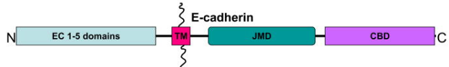
1. E-Cadherin Structure
E-Cadherin structure includes extracellular domain, transmembrane domain and intracellular domain. The extracellular domain contains multiple calcium-binding sites important to adhesion function. Transmembrane domain consists of α-helix, fixing proteins on cell membrane. Intracellular domain connects with cytoskeleton and affects cell adhesion and signal transduction via binding with regulatory proteins. The structure forms calcium-dependent adhesion via some extracellular and adjacent cells, ensuring stable junction and tissue.
2. E-Cadherin Function
Molecular mechanisms of E-Cadherin in cell adhesion include calcium-dependent binding, involvement of intracellular nectins and regulation of signaling pathway. These mechanisms maintain adhesive stability among cells, and are very important to tissue structure and function.
2.1. Intercellular Adhesion
Extracellular domain of E-Cadherin contains several calcium-dependent binding sites, which can bind with E-Cadherin of adjacent cells to form transmembrane junction and promote cell adhesion. The intracellular domain interacts with cytoskeletal proteins(e.g. α-catenin and β-catenin) to improve adhesive stability among cells. Besides, E-Cadherin also binds with signal transduction protein(β-catenin) to regulate cell proliferation, migration and apoptosis, affecting intercellular adhesive stability.
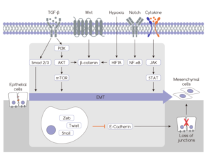
2.2. E-Cadherin and EMT
Epithelial-mesenchymal transition(EMT) is a gradual process, involved in the phenotypic transition of epithelial cells to interstitial cells. In tumor, EMT enables the metastatic ability of cancer cells, showing reassembly of the cytoskeleton and improved movement and invasion. Due to down-regulation of E-Cadherin, cells lose epithelial features but obtain interstitial features, resulting in morphological change and increased migration. Down-regulation of E-Cadherin is achieved via transcription regulation and protein degradation, and plays an important role in physiological and pathological processes(e.g. embryonic development, tumor metastasis and tissue repair etc).
2.3. Regulation of Cell Differentiation
E-Cadherin promotes cell polarity and differentiation via binding between extracellular domain and adjacent cells. Besides, the interaction between E-Cadherin and β-catenin affects Wnt signaling pathway and regulates cell fate determination(e.g. differentiation of stem cell to specific cell line).
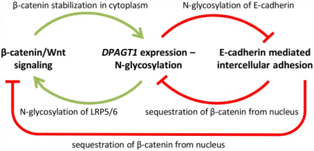
3. E-Cadherin and Wnt pathway
Cellular network consists of conserved metabolic pathways, including N-glycosylation, Wnt/β-catenin signaling pathway and E-Cadherin mediated intercellular adhesion etc. These pathways play an important role in balancing cell proliferation and intercellular adhesion and maintaining homeostasis of differentiated tissues. The regulator β-catenin shared by these pathways is not only the structural component for E-Cadherin connection, but also the co-transcription activator of Wnt pathway.
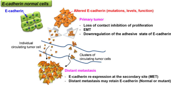
4. Role of E-Cadherin in Cancer Cell
Researches show invasion and metastasis of cancer cells are closely related to deficiency of intercellular adhesion protein. Especially, deficiency of E-Cadherin promotes cancer cell metastasis. Cancer cells are dissociated from primary tumor via EMT process, invade peripheral tissues and migration. However, E-Cadherin is still expressed in some invasive cancers(e.g. infiltrating ductal breast cancer). Deficiency may decrease cell viability and metastatic ability. Thus, role of E-Cadherin in cancer cell is very complex, dependent on types and environment of cancer cell.
5. Antibody‒drug Conjugates(ADCs)
pCAD x CDH17 bispecific antibodies developed by NOVARTIS show obvious anti-tumor activity and provide new method for cancer treatment, especially important to overcome tumor heterogeneity.
On Jan 2nd, 2025, SIM0505-CDH6(ADC) developed by Simcere was approved by clinical trials, mainly focusing on advanced solid tumor(e.g. especially for ovarian cancer and kidney cancer).
6. Application of CDH Family in CAR-T Therapy
On Oct 31st 2023, the first CAR-T cell therapy targeting CDH17(CHM 2101) was approved by FDA. Early researches show the therapy in tumor heterotransplantation and mouse model can completely remove CDH17 expressed by neuroendocrine tumor and gastrointestinal cancer cells. CHM 2101 is safer than other targeted therapy, and non-toxic to normal intestinal epithelial cells. The treated murine small intestine and colon are not injured. Structural damage is not obvious in stomach, pancreas, heart, liver and kidney, showing CDH17 may be a safer solid tumor target.
7. Recommended Products
| Recommended Proteins | |||
| Cat.No | Product Name | Host | Mol. Weight |
| Pr22006 | Recombinant Human CDH1 | Mammalian Cells | 70&85 kDa |
| P1165 | Recombinant Human CDH2 | E.Coli | 35 kDa |
| Pr22167 | Recombinant Human CDH3 | Mammalian Cells | 88-110 kDa |
| P4924 | Recombinant Human CDH3 | E.Coli | 60.1 kDa |
| Pr22721 | Recombinant Human CDH6 | Mammalian Cells | 110-130 kDa |
| Pr22722 | Recombinant Human CDH6 | Mammalian Cells | 109 kDa |
| Pr22723 | Recombinant Human CDH8 | Mammalian Cells | 89 kDa |
| P5398 | Recombinant Human CDH11 | E.Coli | 63.8 kDa |
| Pr23080 | Recombinant Human CDH11 | Mammalian Cells | 110 kDa |
| Pr23081 | Recombinant Human CDH11 | Mammalian Cells | 82 kDa |
| P1156 | Recombinant Human CDH13 | E.Coli | 28.7 kDa |
| P5399 | Recombinant Human CDH13 | E.Coli | 60.9 kDa |
| P5400 | Recombinant Human CDH15 | E.Coli | 20.4 kDa |
| Pr22331 | Recombinant Human CDH16 | Mammalian Cells | 90-115 kDa |
| Pr22199 | Recombinant Human CDH17 | Mammalian Cells | 110-130 kDa |
| P5751 | Recombinant Human CDH18 | E.Coli | 60.9 kDa |
| Pr22471 | Recombinant Mouse CDH3 | Mammalian Cells | 120 kDa |
| Recommended Antibodies | |||
| Cat.No | Product Name | Host | Clonality |
| FNab10112 | CDH1 antibody | Mouse | monoclonal |
| FNab02617 | CDH1 antibody | Rabbit | polyclonal |
| FNab02618 | CDH1 antibody | Rabbit | polyclonal |
| FNab05571 | CDH2 antibody | Mouse | monoclonal |
| FNab05569 | CDH2 antibody | Rabbit | polyclonal |
| FNab05570 | CDH2 antibody | Rabbit | polyclonal |
| FNab10068 | CDH2 antibody | Rabbit | polyclonal |
| FNab06187 | CDH3 antibody | Rabbit | polyclonal |
| FNab10067 | CDH3 antibody | Rabbit | polyclonal |
| FNab07200 | CDH4 antibody | Rabbit | polyclonal |
| FNab10907 | CDH5 antibody | Rabbit | polyclonal |
| FNab10805 | CDH6 antibody | Rabbit | polyclonal |
| FNab01188 | CDH7 antibody | Rabbit | polyclonal |
| FNab01546 | CDH10-specific antibody | Rabbit | polyclonal |
| FNab01184 | CDH13 antibody | Rabbit | polyclonal |
| FNab01185 | CDH16 antibody | Rabbit | polyclonal |
| FNab01547 | CDH17 antibody | Rabbit | polyclonal |
| FNab01186 | CDH18 antibody | Rabbit | polyclonal |
| FNab01187 | CDH20 antibody | Rabbit | polyclonal |
| FNab01548 | CDH23 antibody | Rabbit | polyclonal |
| FNab01549 | CDH26 antibody | Rabbit | polyclonal |
REFERENCES
[1]E-Cadherin Induces Serine Synthesis to Support Progression and Metastasis of Breast Cancer, PMID: 38959339.
[2]E-Cadherin Mutational Landscape and Outcomes in Breast Invasive Lobular Carcinoma, PMID: 39025406.