Abstract: EGFR(Epidermal Growth Factor Receptor) is a HER family member, and widely distributed on the surface of various cells. Roles of EGFR include regulation of cell growth, proliferation and differentiation. The mutation or overexpression is closely related to tumor. In recent years, EGFR researches focus on small molecule inhibitors, ADC drugs and polyclonal and monoclonal antibodies. The drug resistance gradually appears. EGFR western blot protocol covering sample preparation, antibody selection and cellular localization is specified below.
Keywords: EGFR(Epidermal Growth Factor Receptor), Western Blot, Sample Preparation, Antibody Selection, Cellular Localization
1. EGFR Western Blot
1.1. Sample Preparation
EGFR is a transmembrane protein and suggested to extract with SDS or strong RIPA lysis buffer. Ensure complete dissolution with ultrasonic treatment. During sample selection, confirm the expression level of EGFR. Although tumor cells usually express, differences still exist. A431 cell(or HeLa cell) can be used as control for high expression. Besides, add sufficient reductants(e.g. unstable DTT or BME) to break disulfide bond during sample preparation. Ensure effective extraction and analysis of EGFR.
1.2. Antibody Selection
It's very important to study phosphorylation of EGFR. Before finding signal peptide, phosphorylation sites and relevant amino acids of EGFR were first reported. Thereafter, signal peptide on the N terminal contains 24 amino acids, and is removed by enzyme after the transport of EGFR to membranes. Researchers usually follow the first reported phosphorylation sites to keep consistency, also explaining differences of phosphorylation sites. Thus, we should remove 24 amino acids to search for relevant antibodies based on UniProt phosphorylation sites.
1.3. Cellular Localization
EGFR is usually located on the surface of cell membrane. Under specific conditions, EGFR can be transported to nucleus and activate transcriptional activity via endocytosis. During WB, nucleoprotein can be reserved. Alternatively, lyse whole-cell proteins completely. During immunofluorescence localization assay, the positive staining of EGFR in nucleus shouldn’t be considered as non-specific staining. This may show the import of EGFR to nuclei. Intranuclear EGFR is one of the important reasons for drug resistance of EGFR inhibitors.
In EGFR related researches, the expression abundance, recognized antibody sites and experimental process affect whether WB can be smoothly performed.
2. Extended Reading
2.1. EGFR Structure and Function
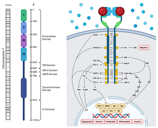
EGFR gene is the tyrosine kinase receptor located on the short arm of chromosome 7, including 28 exons. As a member of ErbB family, the receptor consists of extracellular domain, transmembrane domain and intracellular domain.
2.2. EGFR Signaling Pathway
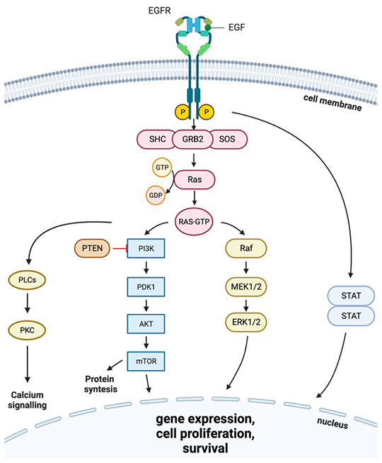
After binding with ligands(e.g. EGF or TNF-α), EGFR forms homo-or heterodimers and activates the activity of tyrosine kinase to initiate autophosphorylation reaction. Activated downstream signaling pathways include Ras/Raf/MEK/ERK1/2, JAK/STAT and PI3K/Akt pathways, regulating cell proliferation, cell cycle, survival respectively via apoptosis inhibition. Tyrosine kinase activity of EGFR is affected by carcinogenic factors(e.g. EGFR gene mutation, increased gene copy number, overexpression etc). Besides, EGFR interacts with integrin pathway to activate matrix metalloproteinase, and also accelerates tumor development via promoting movement, invasion and metastasis.
3. Recommended Products
| Recommended Recombinant Proteins | |
| Cat.No | Product Name |
| P0611 | Recombinant Human EGFR |
| Pr22252 | Recombinant Human EGFR vIII |
| Recommended Antibodies | |
| Cat.No | Product Name |
| FNab10146 | EGFR antibody |
| FNab02669 | EGFR-Specific antibody |
| Recommended ELISA Kits | |
| Cat.No | Product Name |
| EH20023 | Human EGFR ELISA Kit |
| QT-EH20023 | Human EGFR QuickTest ELISA Kit |
| EM1602 | Mouse EGFR ELISA Kit |
| QT-EM1602 | Mouse EGFR QuickTest ELISA Kit |
REFERENCES
[1]Glutamate dehydrogenase 1-catalytic glutaminolysis feedback activates EGFR/PI3K/AKT pathway and reprograms glioblastoma metabolism, PMID: 39446525.
[2]Emodin Promotes Peripheral Nerve Repair by Modulating Inflammasome Activation Through Autophagy via the EGFR/PI3K/AKT/mTOR Pathway, PMID: 40135386.