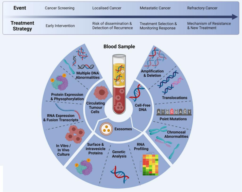
Abstract: Blood tumor markers are biomolecules detecting and monitoring the existence and development of certain types of cancer. They usually evaluate concentration of specific substances in the body via detecting blood sample. The level of these substances may increase when suffering from some tumors. The increased level of markers is not induced by all tumors, and reflects the existence of tumor. Markers are applied in the early detection and diagnosis as well as surveillance of therapeutic effects.
Keywords: Blood Tumor Markers, Oncofetal Antigen, Cancer Treatment and Diagnosis, Therapeutic Monitoring, Prognostic Evaluation
1. Oncofetal Antigen Tumor Markers
Tumor biomarkers like AFP and CEA found in 1960 have been widely used till now. They are oncofetal antigens, which appears during fetal period and disappears in adulthood. The reproduction of these oncofetal antigens in cancer patients may be related to the activation of some genes disabled in adulthood. These genes can produce oncofetal antigens during transformation of malignant cells. Currently, only a few oncofetal antigens tumor markers play an important role in clinical cancer treatment.
1.1. AFP
AFP(Alpha-fetoprotein) found in 1956 is the single-chain glycoprotein containing 3-5% carbohydrate. It's encoded by AFP gene on Chromosome 4. AFP is the main circulating protein during fetal period. The concentration gradually decreases after 18 months of birth. AFP in healthy adult serum is usually lower than 10μg/L. AFP is the most common tumor marker in hepatocellular carcinoma(HCC). AFP level in about 80% HCC patients increases, and are especially screened in China and Japan. AFP plays an important role in diagnosis of HCC. AFP level of about 40% early patients is normal, and may increase in chronic liver disease.
1.2. CEA
Carcinoembryonic antigen(CEA) was extracted from human colorectal cancer and embryonic tissue in 1965, and belongs to glycoprotein at the cell surface. The gene is located on Chromosome 19. The production of CEA in digestive tract starts from early fetal period, and is highly expressed in various tumors. As the broad-spectrum tumor biomarker, CEA level in cancer patients increases, including 70% colorectal cancer, 55% pancreatic cancer. CEA level is proportional to tumor burden, and usually used in diagnosis, prognosis and recurrence monitoring. Due to the lower sensitivity and specificity, CEA has to be used with other markers.

2. Assisted Diagnostic Markers
2.1. PSA
Prostate-specific antigen(PSA) is the enzyme secreted by prostatic epithelial cells and encoded by prostate-specific gene Kallikrein 3. PSA was first found in 1970. The increase of PSA level in serum is relevant to prostatitis, benign prostatic hyperplasia and prostate cancer. Positive cut-off value of PSA for early diagnosis of prostate cancer is 10 ng/mL above. PSA is widely applied in cancer detection and patient management. The lower specificity of 20 - 40% can’t distinguish inactive and invasive cancer, resulting in the overdiagnosis of prostate cancer.
2.2. NSE
Neuron-specific enolase(NSE) is the enzyme in neurons and peripheral neuroendocrine cells. NSE was recognized in 1965. The increase of NSE is relevant to tumor transforming, and plays an important role in diagnosis and prognosis of cancer derived from nerve and neuroendocrine. In small cell lung cancer(SCLC), NSE is the biomarker for staging and monitoring treatment. Besides, high level of NSE in NSCLC patients is related to short progression-free survival and overall survival. Thus, NSE is the important marker for cancer treatment prediction and independent prognostic factor.
3. Therapeutic Monitoring Markers
3.1. CA72-4
Carbohydrate antigen 72-4(CA72-4) is the mucin embryonic antigen found in 1981 and mainly exists in human adenocarcinoma. The increased serum level is the effective diagnostic indicator for various cancers(e.g. gastric cancer, pancreatic cancer, colorectal cancer). The sensitivity and specificity for gastric cancer are 49% and 96% respectively, higher than other tumor markers. However, higher expression of CA72-4 in normal tissues may induce false-positive, especially for atrophic gastritis patients. Thus, other biomarkers are required for improving the sensitivity and specificity of diagnosis. Above all, CA72-4 plays an important role in screening and therapeutic effect assessment of gastric cancer.
3.2. CA125
CA125 is a highly glycosylated mucin, first found in ovarian cancer cell line OVCA433 via monoclonal antibody OC125. CA125 is the important biomarker for monitoring epithelial ovarian cancer. The diagnostic sensitivity is about 70%. CA125 level is related to the development and recurrence of ovarian cancer. 35U/mL below is considered to be effective for postoperative or chemotherapeutic CA125 level in the serum. 70U/mL or above may indicate the possible recurrence. Besides, CA125 can be also applied in the diagnosis of other non-ovarian cancers(e.g. cervical and gastric cancer). CA125 may increase in about 1% healthy people and 5% patients with benign disease.
3.3. TPS
Tissue polypeptide specific antigen(TPS) is the M3 antigen on cytokeratin, mainly synthesized in S and G2 stage. Serum level of TPS reflects cell proliferation activity, and is related to the number of cells in proliferation stage instead of total number of tumor cells. The obvious increase of TPS level in various cancers (e.g. endometrial cancer, bladder cancer, non-small cell lung cancer etc) can monitor therapeutic effect and predict tumor recurrence. TPS can also predict prognosis in breast and gastric cancer. Although the sensitivity is limited, TPS still plays an important role in cancer management.
4. Prognostic Evaluation Markers
4.1. SCCA
Squamous cell carcinoma antigen(SCCA) is the tumor specific antigen first separated from cervical squamous cell carcinoma tissues in 1970. SCCA is applied in diagnosis of cervical cancer. The serum level of SCCA is related to tumor stage, invasion depth and recurrent risk. High level of SCCA patients has the higher mortality risk. Researches show SCCA increases in oral cancer, esophageal cancer, lung cancer. Although the sensitivity is limited, SCCA is still the important biomarker for cancer diagnosis and prognosis, non-small cell lung cancer monitoring and chemotherapeutic efficacy evaluation.
4.2. AFU
Alpha-l-fucosidase(AFU) consists of AFU1 and AFU2, encoded by FUCA1 and FUCA2 respectively. AFU is the lysosomal enzyme without alpha-l-furanose residue on the glycoprotein terminal. AFU is involved in metabolism of glycoprotein and glycolipid, and widely distributed in human tissue and blood. Usually, AFU level in the serum is low but rapidly increase due to tumor stage and size. Researches show AFU is the important biomarker for hepatocellular carcinoma(HCC), and can diagnose HCC six months earlier than ultrasonic examination. The level is very important for prognosis and recurrent risk evaluation.
4.3. LDH
Lactic dehydrogenase(LDH) is the enzyme catalyzing reversible conversion between pyruvic acid and lactic acid. LDH consists of LDHA and LDHB. LDHA is expressed in most tumor tissues, and closely related to cancer cell proliferation, increased invasion, metastasis and resistance in chemotherapy. Researches show increased LDH level in serum is related to poor prognosis of various cancers(especially melanoma, renal cell carcinoma and colorectal cancer). LDH is considered to be the important biomarker for prognosis, contributing to individual treatment guidance and evaluation for diagnosis and therapeutic effect of cancer.
5. Conclusion
Blood tumor markers are important tools for cancer diagnosis, prognostic evaluation and therapeutic monitoring. Specific concentration of these markers is usually found in patients’ serum, showing the existence of tumor and biological properties.
6. Recommended Products
REFERENCES
[1] Use of tumor markers in distinguishing lung adenocarcinoma-associated malignant pleural effusion from tuberculous pleural effusion, PMID: 38583522.
[2] Preoperative inflammatory markers and tumor markers in predicting lymphatic metastasis and postoperative complications in colorectal cancer: a retrospective study, PMID: 39966788.
