Abstract: Mitochondria are organelles carrying unique genetic codes in eukaryotic cells. mtDNA encodes 13 proteins which are key components of OXPHOS complex. Mitochondria manage to produce energy required by most cells via oxidative phosphorylation, and are also involved in several important biological processes, such as regulation of apoptosis, storage and regulation of calcium ion, production and regulation of reactive oxygen species(ROS). Thus, mitochondria play an important role in inflammatory response. Mitochondrial dysfunction and inflammation interact with each other.
Keywords: Mitochondrial Dysfunction, Inflammation, Mitochondrial Disease Diagnosis, Targeted Therapy
1. Role of Mitochondria in Inflammatory Response
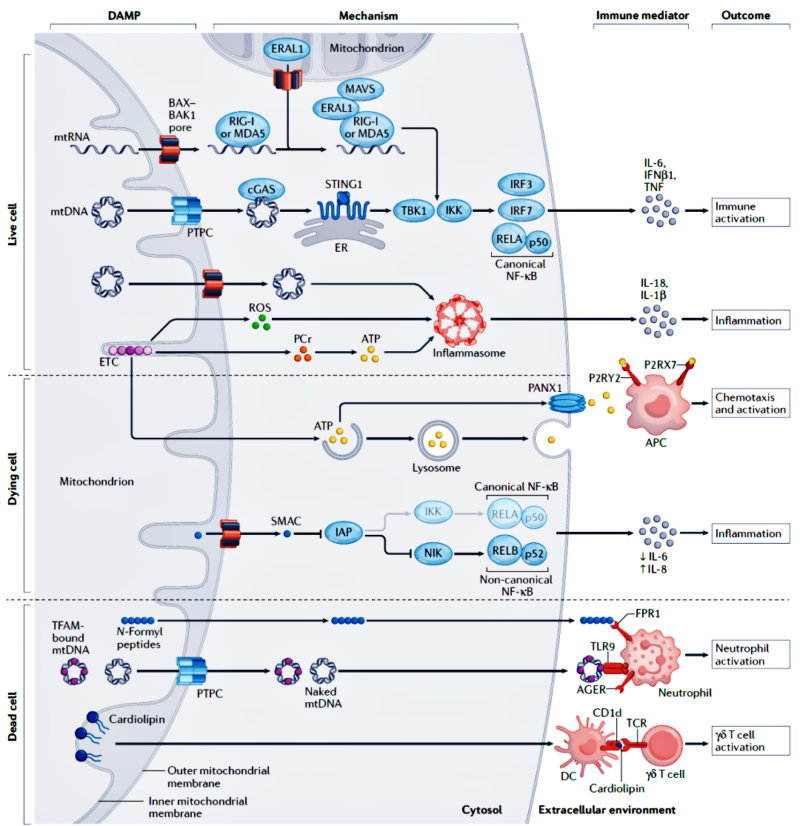
When mitochondria are damaged by factors like infection, oxidative stress or toxin etc, internal components may be leaked into cytoplasm to induce inflammatory response. Without the protection of histone, damaged mtDNA escapes to cytoplasm more easily. mtDNA is rich in unmethylated CpG island. These sequences can be recognized by intracellular pattern recognition receptors(e.g. TLR9). The recognition process can activate the specific inflammatory signaling pathway, and promote cells to produce various pro-inflammatory cytokines(e.g. TNF-α, IL-1β), which cause or enhance inflammatory response. Thus, mitochondrial damage affects normal cell function, and is involved in inflammatory mechanism.
2. Regulation of Mitochondria in Inflammatory Signal
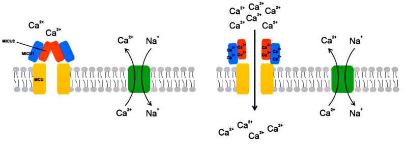
Mitochondria are one of the main producers for ROS. Appropriate amount of ROS plays an important role in regulating cellular physiological functions, including regulation for inflammatory response. In immunocytes, lower level of ROS can activate specific signaling pathway, and promote cell activation and secretion of inflammatory factors(e.g. cytokines). However, more generated ROS can cause oxidative stress, which can enhance inflammatory response by damaging cells. Besides, mitochondria are very important for maintaining balanced concentration of calcium ion in the cell. Calcium ion is the key intracellular signaling molecule, and plays an important role in inflammatory process. Mitochondria helps to regulate homeostasis of calcium ion in the cell via absorbing and releasing calcium ion. Mitochondrial dysfunction may cause homeostasis imbalance of calcium ion, and further affects inflammatory signal transduction as well as performance of cellular immune function.
3. Cause of Mitochondrial Disease
3.1. Role of Mitochondria in Cancer
Apoptosis is the protection mechanism for inhibiting tumor growth via killing tumor cells. One of the main inducing factors is the increased ROS level caused by increased mitochondrial metabolism. Excessive ROS can damage mitochondrial function, resulting in depolarization of mitochondrial membrane and activation of apoptotic pathway. Besides, mitochondria play the key role in immune evasion of tumor cells via promoting or inhibiting the process, and also affect complex mechanism of immune response.
3.2. Mitochondria and Heart Disease
Mitochondria associated programmed cell death is the key regulatory factor related to various cardiovascular diseases(e.g. heart failure, atherosclerosis, aneurysm etc). Cardiomyocyte apoptosis always occurs with congestive heart failure. Untimely clearance for apoptosis of endothelial cells can promote the formation of atherosclerosis. Besides, ischemia/reperfusion(I/R) or oxidative stress-induced necrosis can also cause heart failure.
3.3. Mitochondrial Metabolic Disease
In type 2 diabetes, glucotoxicity, lipotoxicity and aggregation of islet amyloid polypeptide cause dysfunction of β cell, and activate ER stress signalling pathways. Then, the level of CHOP(pro-apoptotic factor) further increases to promote the production of ROS, mitochondrial dysfunction and apoptosis.
4. Mitochondria Targeted Therapy
There are various treatment methods for targeted mitochondria against infections:
- Improve mitochondrial function by using antioxidants and coenzyme Q10;
- Decrease oxidative stress and improve the resistance of cells against infections;
- Regulate ROS level in mitochondria to promote immune activation without causing over-oxidative stress;
- Intervene to absorb and release calcium ion via mitochondria, maintain normal concentration of calcium ion in the cell, prevent abnormal immune response;
- Develop inhibitors for recognizing mtDNA like pattern recognition receptors(TLR9) to prevent activation of inflammatory signaling pathway;
- Promote selective degradation of damaged mitochondria(i.e. mitophagy) to remove unhealthy mitochondria which may cause chronic inflammation;
- Explore small molecule inhibitor which directly affects interactions between virus and mitochondria, alternatively regulate mitochondria induced apoptotic pathway to restrict virus replication;
- Improve metabolism state of mitochondria by specific diet or supplement.
These strategies are currently in the research stage. With the deep understanding of mitochondrial biology and pathology, more effective treatment methods with less side effect will be developed in the future.
5. Mitochondrial Disease Diagnosis
Mitochondrial disease diagnosis processes include clinical features, biochemical metabolism and imaging examination etc. Highly suspected mitochondrial diseases are required for gene detection. When the gene detection is negative, clinical evaluation should be conducted again. If the mitochondrial disease is still suspected, muscle or skin biopsy is required. Pathological analysis and enzymatic assay of respiratory chain are also conducted. Meanwhile, genetic analysis for tissue sample is necessary.
6. Recommended Products
|
Recommended Proteins |
|||
|
Cat.No |
Product Name |
Mol. Weight |
Host |
|
Recombinant Human SOD2 |
24 kDa |
E.Coli |
|
|
Recombinant Human TFAM |
22.3 kDa |
E.Coli |
|
|
Recombinant Human ATP5F1A |
75.8 kDa |
E.Coli |
|
|
Recombinant Human SDHA |
42.4 kDa |
E.Coli |
|
|
Recombinant Hamster TNFA |
29.3 kDa |
E.Coli |
|
|
Recommended Antibodies |
|||
|
Cat.No |
Product Name |
Tested Application |
Antibody Type |
|
ATP5F1A antibody |
ELISA, IHC, WB, IF, IP |
Rabbit pAb |
|
|
COX4I1 antibody |
ELISA, WB, IHC, IF, IP, FC |
Rabbit pAb |
|
|
POLG2 antibody |
ELISA, IHC, WB, IF |
Rabbit pAb |
|
|
SDHA antibody |
ELISA, WB, IHC, IF |
Rabbit pAb |
|
|
SOD2 antibody |
ELISA, WB, IF, IHC, IP |
Rabbit pAb |
|
|
TFAM antibody |
ELISA, WB, IHC, IP |
Rabbit pAb |
|
|
UCP2 antibody |
ELISA, WB, IHC |
Rabbit pAb |
|
|
Recommended ELISA Kits |
|||
|
Cat.No |
Product Name |
Range |
Sensitivity |
|
Human TFAM QuickTest ELISA Kit |
31.25-2000pg/ml |
18.75pg/ml |
|
|
Human TNF-α QuickTest ELISA Kit |
15.625-1000pg/ml |
9.375pg/ml |
|
|
Mouse TNF-α QuickTest ELISA Kit |
3.906-250pg/ml |
2.344pg/ml |
|
|
Rat TNF-α QuickTest ELISA Kit |
3.906-250pg/ml |
2.344pg/ml |
|
|
Human IL-1β QuickTest ELISA Kit |
3.906-250pg/ml |
2.344pg/ml |
|
|
Mouse IL-1β QuickTest ELISA Kit |
31.25-200pg/ml |
1.875pg/ml |
|
|
Rat IL-1β QuickTest ELISA Kit |
31.25-2000pg/ml |
18.75pg/ml |
|
|
Human SDH ELISA Kit |
0.313-20ng/ml |
0.188ng/ml |
|
|
Rat SDH ELISA Kit |
31.25-2000pg/ml |
18.75pg/ml |
|
|
Human SOD2 ELISA Kit |
0.313-20ng/ml |
0.188ng/ml |
|
|
Mouse SOD2 ELISA Kit |
0.313-20ng/ml |
0.188ng/ml |
|
|
Rat SOD2 ELISA Kit |
31.25-2000pg/ml |
18.75pg/ml |
|
|
Mouse TFAM ELISA Kit |
31.2-2000pg/ml |
18.75pg/ml |
|
|
Human COX4I1 ELISA Kit |
0.156-10ng/ml |
0.094ng/ml |
|
|
Rat Cox4i1 ELISA Kit |
0.156-10ng/ml |
0.094ng/ml |
|
|
Human UCP2 ELISA Kit |
0.156-10ng/ml |
0.094ng/ml |
|
REFERENCES
[1]Senescent glia link mitochondrial dysfunction and lipid accumulation, PMID: 38839958.
[2]CKLF induces microglial activation via triggering defective mitophagy and mitochondrial dysfunction, PMID: 37908119.