Abstract: Peroxidase-labeled antibody method was introduced in 1968. For most clinical researches, it's the first actual application for antibodies in paraffin embedded tissues(Nakane, 1968). In fact, each IgG molecular connectivity of traditional HRP conjugated IgG secondary antibody is limited to 3 HRP moleculars. The mole ratio is sufficient in regular WB or ELISA. However, the sensitivity for detection samples between low pg and fg may be insufficient. Signal amplification step is not required for IHC.
Keywords: HRP Secondary Antibody, Peroxidase Antibody, IHC
1. Immunohistochemical Methods
In the past decades, improvements for IHC reagents and methods make the sensitivity of detection system increased. The antigen detection ability of PAP and APAAP (Sternberger et al. 1970) is higher than standard ABC/SABC and labeled streptavidin biotin. The LSAB method can be obtained by the chain polymer conjugation technique developed in the last decade of the last century.
However, PAP and APAAP methods appear to produce spatial barrier, which forms a huge skeleton structure in the aqueous environment. As a result,the reagent penetration is decreased, especially for hidden nuclear antigen.
Biotins(Vitamin H) contained in many tissues have been introduced by ABC(SABC) method. Hence, they can bind with ABC(SABC). Thus, non-specific staining occurs.
2. FineTest Poly HRP Secondary Antibody
In order to enhance signal and reduce non-specific reaction, Fine Biotech adopts new polymerase labeling system, including poly-HRP conjugated Goat anti Rabbit IgG and poly-HRP conjugated Goat anti Mouse IgG. More enzymes will be connected with single antibody by the arm structure. Meanwhile, the best structure of the new system enhances the signal during immunochemical process and reduces experimental steps.
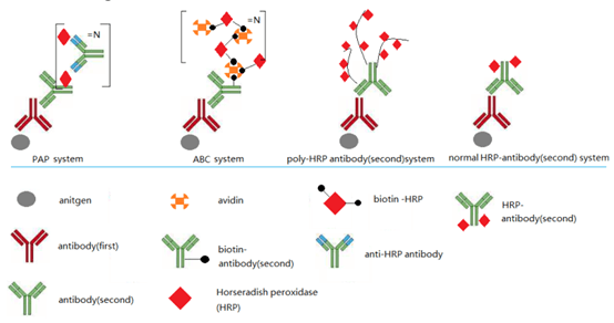
Figure 1. schematic diagram of detection principle.
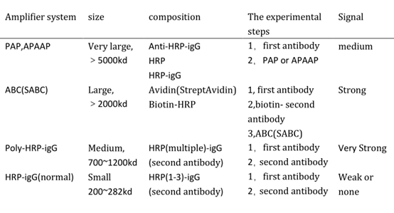
Table 1. Data Comparison between Different Detection System.
3. IHC Test Using Poly HRP Secondary Antibody
Two kinds of FineTest antibodies with high and low level targets are tested, using two secondary antibodies: poly-HRP conjugated Goat anti Rabbit IgG(ES-0001) and poly-HRP conjugated Goat anti Mouse IgG(ES-0002). DAB substrate kit also comes from FineTest(IHC0005).
Table 2. Comparison between High and Low Level Targets.
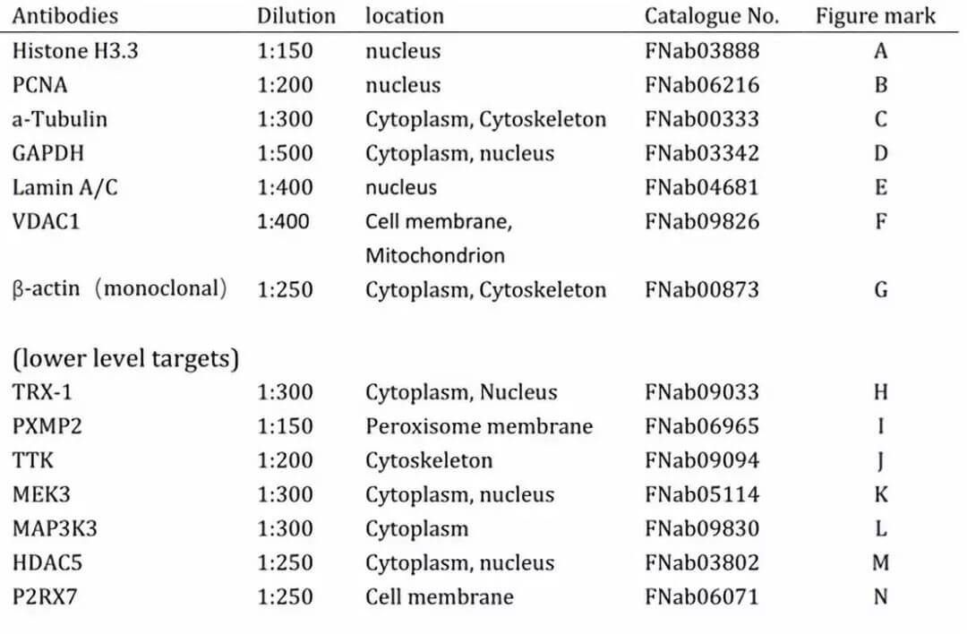
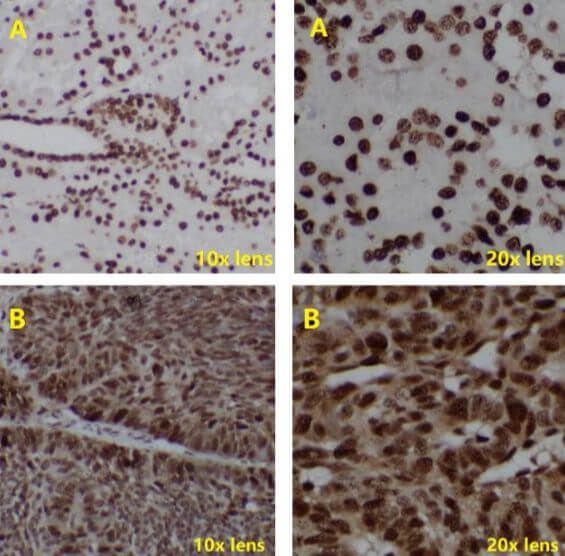
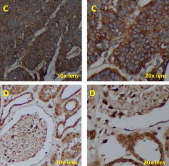
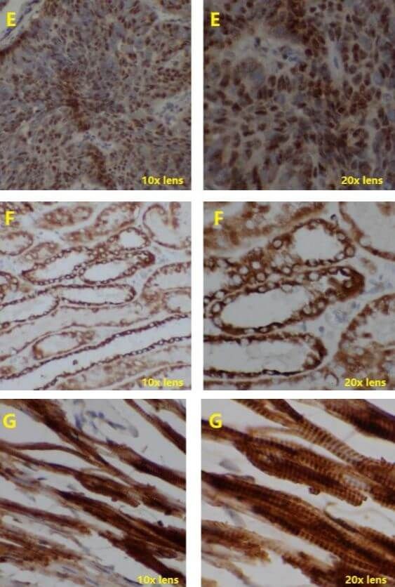
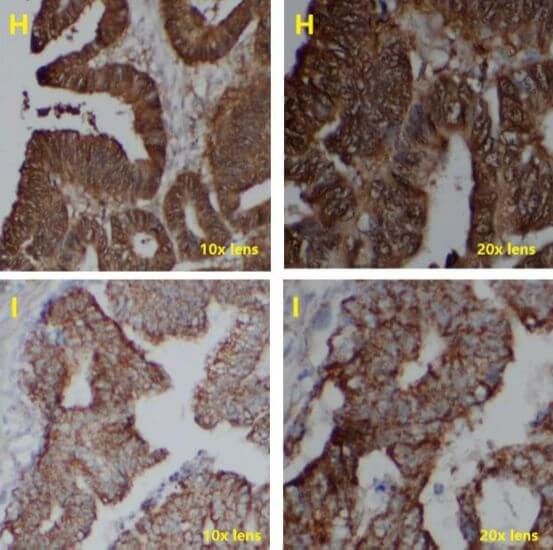


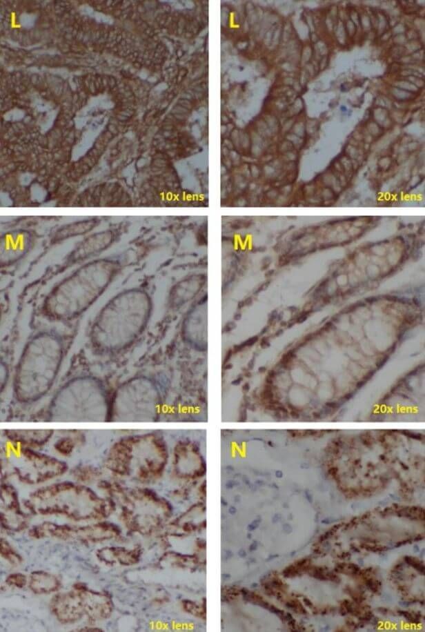
Figure 2. Immunostaining tissue samples: human kidney(A,D,F,N); human liver cancer(B,E); human breast cancer(C,J,K); human colon cancer(H,I,L,M); rat heart(G; Dilution ratio for poly-HRP conjugated Goat anti Rabbit IgG and poly-HRP conjugated Goat anti Mouse IgG: 1:100.
4. Negative Control Test
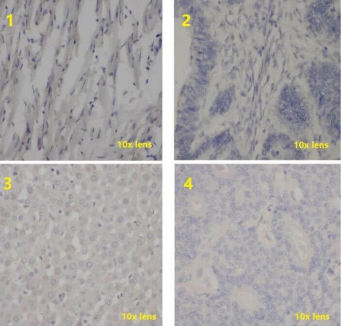
Figure 3. Negative control test for immunostaining tissue samples: rat heart(1); human colon tumor(2); rat liver(3); human breast tumor(4); the serum was negative primary antibody; Dilution ratio for poly-HRP conjugated Goat anti Rabbit IgG and poly-HRP conjugated Goat anti Mouse IgG: 1:50; Staining for 10min with DAB kit; nucleus is stained by hematoxylin.
5. Conclusion
Poly-HRP conjugates have excellent signal amplification performance and don’t produce non-specific results. Furthermore, the experiment operations are greatly simplified.
REFERENCES
Improving the sensitivity of traditional Western blotting via Streptavidin containing Poly-horseradish peroxidase (PolyHRP), PMID: 31021001.