Abstract: Cells in flow cytometry experiments are processed as single cell suspension, following surface marker staining or intracellular target staining for further analysis or sorting through fixation and permeabilization. Important roles of flow cytometry buffers for sample preparation include protection of antigenic epitopes, ensuing antibody-target binding and reduced interference of background signal. Thus, selection of suitable FACS buffers are very important for accurate and reliable experimental results. Besides, optimization is required for detailed experimental requirements.
Keywords: Flow Cytometry Buffers, FACS Buffer, Staining Buffer, Blocking Buffer, Fixation Buffer, Sorting Buffer, Permeabilization Buffer
1. Role of Flow Cytometry Buffers
Applications of flow cytometry buffers include cell protection, optimization of staining effects, decrease of non-specific binding, improvement of signal stability, assistance to cell fixation, permeabilization and sorting, stable chromophore, improved accuracy and data quality.
2. Staining Buffer
Staining buffer is based on phosphate buffer(PBS). Usually, Ca²⁺ and Mg²⁺ are not found in PBS to prevent cell aggregation. 0.1-1% BSA or 1-10% FBS is usually added into the buffer to reduce the non-specific binding of antibody or fluorescent dye via blocking non-specific binding sites. Commercial staining buffers usually contain 0.1-1% NaN₃ as preservatives. Besides, staining buffer can be also applied in washing during antibody dilution and staining.
3. Cell Processing
3.1. Cell Sorting Buffer
Sorting buffers usually use 1xPBS or HBSS without calcium and magnesium ion. 1-5% FBS or 0.1-1% BSA is also added. Serum concentration should not be too high, and avoid to affect sorting results. Culture mediums containing phenol red are not suggested, since phenol red may interfere with sorting. Add 25mM HEPES into the buffer to maintain pH stability. Add 2-5mM EDTA to prevent aggregation of easily adhesive cells. If there are many dead cells, add 25-50µg/ml DNAse and 1-5mM MgCl₂. Cell collection solution can use 1xPBS/ HBSS(with FBS/BSA) or culture medium containing serum. Antibiotics can be added to prevent contamination.
3.2. RBC Lysis Buffer
Whole blood and tissue homogenate usually contain a large number of red blood cells, affecting collection and detection of target cells. Thus, during preparation of single cell suspension(e.g. peripheral blood or mouse spleen samples), RBC lysis buffer is usually used to remove red blood cells, following further staining and detection. Main component of RBC lysis buffer is ammonium chloride, lysing red blood cells with osmotic pressure difference principle. RBC lysis buffers in the market are usually 1x or 10x working solutions, and properly diluted according to the manual.
3.3. Fixation and Permeabilization Buffer
Fixation and permeabilization buffers are applied in intracellular antigen staining, aiming to fix cell structure and permeate cell membrane. Then, antibodies can enter cells to bind with the target antigen. Fixation steps usually use 1-4% formaldehyde(soluble in PBS). Incubate for 10-15min at room temperature to keep cell morphology and antigen integrity, and lock intracellular protein structure. Then, process cells with proper permeabilization buffers. Common buffers include Triton X-100 and saponin etc, and can permeate cell membrane via allowing the entry of antibodies or dyes in+to cells. Selection of proper permeabilization buffers depend on experimental requirements.
Experimental results for two kinds of fixation and permeabilization buffers(K082R, K081) are shown below.
Case 1: TH1 Cell Detection in Mouse Spleen
| Target | Fluorescein | Clone ID | Cat.No |
|---|---|---|---|
| CD3 | FineTest®647 | 17A2 | F647-30127 |
| CD4 | FITC | RM4-5 | FITC-30128 |
| IFN-γ | PE | XMG1.2 | PE-30074 |
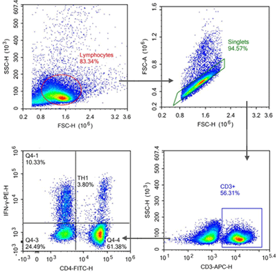
TIPS:
① Obvious grouping for CD3/CD4 without isotype control. IFN-γ detection requires for isotype control.
② Stimulation of PMA can cause endocytosis of CD4+T cell epitope. CD4 antibody(Clone ID: RM4-5) can reduce endocytic effects of CD4 epitope via detecting permeabilized CD4 epitope. Grouping of CD4 cell is clearer.
③ As the cytokine, IFN-γ should be detected after blocking, fixation and permeabilization.
Case 2: Treg Cell Detection in Mouse Spleen
| Target | Fluorescein | Clone ID | Cat.No |
|---|---|---|---|
| CD4 | FITC | RM4-5 | FITC-30128 |
| CD25 | APC | PC-61.5.3 | APC-30017 |
| FOXP3 | PE | 3G3 | PE-30111 |
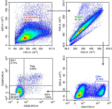
TIPS:
① Obvious grouping for CD4 without isotype control. Detection of CD25 and FOXP3 requires for isotype control.
② FOXP3 can't be detected using improper fixation and permeabilization kit. K081(Foxp3/Transcription Factor Staining Kit) is recommended.
4. Blocking
4.1. Fc Blocker
The surface of some immune cells(especially monocytes, macrophages and NK cells) expresses Fc receptor. These receptors can bind with Fc region of antibodies. Thus, during staining of these cells, commercial Fc block reagents or sera are required for blocking. Fc blocking is performed before immunostaining. Incubate for 5-10min at room temperature to reduce non-specific binding.
4.2. Monocyte Blocker
Monocyte blocker is the special reagent for reducing non-specific binding between monocytes/macrophages and fluorescent dyes. Non-specific binding may happen between some fluorescent dyes(e.g. PE/Dazzle™ 594, PE/Cy5, PE/Cy7, APC/Cy7, APC/Fire™ 750 etc) and monocytes/macrophages, resulting in background noise, false positive signal and inaccurate flow cytometry. Thus, monocyte blocker can effectively reduce interference and ensure reliable experimental results.
5. Dye Processing
5.1. Polymer Dyes Staining Buffer
Polymer dyes staining buffer is especially designed for reducing non-specific binding during staining and interaction among dyes. Thus, the accuracy and stability of staining are improved. If several Brilliant series dyes are used together with polymer dyes(e.g. BV, BUV, SB), Brilliant Stain Buffer or Super Bright Staining Buffer is suggested to add to ensure the best staining effect and avoid interference among dyes.
5.2. Tandem Stabilizer
Tandem dyes(e.g. PE-Cy5, PE-Cy7, APC-Cy7) consist of two fluorophores(donor and receptor). When laser excites donor, photon is transferred to receptor for luminescence via fluorescence resonance energy transfer(FRET). Tandem dyes provide experimental flexibility. But they are easily affected by photobleaching, chemical degradation or broken fluorophores during fixation. Thus, decreased signals or offset compensation affected data analysis. Tandem stabilizer can be added to prevent dye degradation so that fixed cells can be stored for 4 days at 4℃ in the dark. Then, data collection is easier.
6. Recommended Products
| Product Category | Product Name | Cat.No | Size | Applications |
| Erythrocyte Lysis Buffer | 10x ACK Lysis Buffer(without fixative) | K080 | 100mL/200mL/500mL | Lyse the erythrocyte in whole blood |
| Buffer | Cell Staining Buffer | K079 | 100mL/200mL/500mL | Dilution of fluorescent antibody in flow cytometry and cell washing |
| Nuclear Staining | PI Reagent | K116 | 50T/100T/200T/500T | DNA staining in cells |
| 7-AAD Reagent | K115 | 50T/100T/200T/500T | DNA staining in cells | |
| DAPI Reagent | K117 | 50T/100T/200T/500T | DNA staining in cells | |
| Stimulating and Blocking | Cell Stimulation and Protein Transport Inhibitor Kit | K083 | 50T/100T/200T | Stimulate/block cytokines |
| Fixation/Permeabilization | Foxp3/Transcription Factor Staining Kit | K081 | 20T | Fixation and permeabilization kit for staining intranuclear antigens |
| Intracellular Fixation/Permeabilization Buffer Kit (Ready To Use) | K082R | 50T/100T/500T | Fixation and permeabilization kit for intracellular factors | |
| FcR Blocking | Purified Anti-Mouse CD16/32 Antibody | FNab30106 | 100μg | Blocking for mouse sample |
| Purified Anti-Human CD16 Antibody | FNab10997 | 100μg | Blocking for human sample | |
| Viability Dye | Calcein AM/PI Live/Dead Double Staining Kit | FNCK022 | 100T/500T | Distinguish dead or live cells |
| FineTest® Red 780(Viability Dye) | K121 | 100T | Distinguish dead or live cells | |
| FineTest®Violet 510(Viability Dye) | K124 | 100T | Distinguish dead or live cells |
REFERENCES
[1] Optimized flow cytometry protocol for dihydrorhodamine 123-based detection of reactive oxygen species in leukocyte subpopulations in whole blood, PMID: 35760097.
[2] A Novel Sorbitol-Based Flow Cytometry Buffer Is Effective for Genome Size Estimation across a Cypriot Grapevine Collection, PMID: 38475579.