VRK2 antibody

Synonyms:Serine/threonine-protein kinase VRK2|Vaccinia-related kinase 2|VRK2 antibody
Catalogue No.:FNab09452Reactivity:Human, Mouse, Rat
Host:RabbitTested Application:ELISA, WB, IP
Clonality:polyclonalIsotype:IgG
Size Price
100µg Inquiry
Dispatch Time: About 3 working days
  • SPECIFICATIONS
  • FIGURES
  • CONDITIONS
  • FAQS
Product Name
VRK2 antibody
Catalogue No.
FNab09452
Size
100μg
Form
liquid
Purification
Immunogen affinity purified
Purity
≥95% as determined by SDS-PAGE
Clonality
polyclonal
Isotype
IgG
Storage
PBS with 0.02% sodium azide and 50% glycerol pH 7.3, -20℃ for 12 months(Avoid repeated freeze / thaw cycles.)
Immunogen
Immunogen
vaccinia related kinase 2
Alternative Names
Serine/threonine-protein kinase VRK2|Vaccinia-related kinase 2|VRK2 antibody
UniProt ID
Q86Y07
Observed MW
67 kDa
Application
Tested Applications
ELISA, WB, IP
Recommended dilution
WB: 1:500-1:5000;IP: 1:200-1:2000
Validated Images
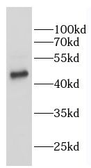
K-562 cells were subjected to SDS PAGE followed by western blot with FNab09452(VRK2 antibody) at dilution of 1:500
Background
Serine/threonine kinase that regulates several signal transduction pathways. Isoform 1 modulates the stress response to hypoxia and cytokines, such as interleukin-1 beta(IL1B) and this is dependent on its interaction with MAPK8IP1, which assembles mitogen-activated protein kinase(MAPK) complexes. Inhibition of signal transmission mediated by the assembly of MAPK8IP1-MAPK complexes reduces JNK phosphorylation and JUN-dependent transcription. Phosphorylates 'Thr-18' of p53/TP53, histone H3, and may also phosphorylate MAPK8IP1. Phosphorylates BANF1 and disrupts its ability to bind DNA and reduces its binding to LEM domain-containing proteins. Downregulates the transactivation of transcription induced by ERBB2, HRAS, BRAF, and MEK1. Blocks the phosphorylation of ERK in response to ERBB2 and HRAS. Can also phosphorylate the following substrates that are commonly used to establish in vitro kinase activity: casein, MBP and histone H2B, but it is not sure that this is physiologically relevant.Isoform 2 phosphorylates 'Thr-18' of p53/TP53, as well as histone H3. Reduces p53/TP53 ubiquitination by MDM2, promotes p53/TP53 acetylation by EP300 and thereby increases p53/TP53 stability and activity.
How many times can antibodies be recycled?

First, usually it's not suggested to recycle antibodies. After use, buffer system of antibodies has changed. The storage condition of recycled antibodies for different customers also varies. Thus, the performance efficiency of recycled antibodies can’t be guaranteed. Besides, FineTest ever conducted the antibody recycling assay. Assay results show recycling times of different antibodies also varies. Usually, higher antibody titer allows more repeated use. Customers can determine based on experimental requirements.

Notes: After incubation, we recycle rest antibodies to centrifuge tube and store at 4℃. High titer antibodies can be stored for a minimum of one week. Reuse about three times.

What are components of FineTest antibody buffer?

Components of FineTest antibody buffer are usually PBS with proclin300 or sodium azide, BSA, 50% glycerol. Common preservative is proclin300 or sodium azide, which is widely applied in the lab and industry.

How about the storage temperature and duration of FineTest antibodies?

Most antibodies are stored at -20℃. Directly-labeled flow cytometry antibodies should be stored at 2 - 8℃. The shelf life is one year. If after sales issues for purchased antibodies appear, return or replacement is available. Usually, antibodies can be still used after the one-year warranty. We can offer technical support services.

Is dilution required for FineTest antibodies? What’s the dilute solution?

Directly-labeled flow cytometry antibodies are ready-to-use without dilution. Other antibodies are usually concentrated. Follow the dilution ratio suggested in the manual. Dilute solution for different experiments also varies. Common antibody dilution buffers are acceptable(e.g. PBST, TBST, antibody blocking buffer).

How to retrieve antibodies for immunohistochemistry?

Common retrieval buffers: Tris-EDTA Buffer(pH 9.0); Citrate Buffer(pH 6.0)

Heat induced antibody retrieval:

Method 1: Water-bath heating: Put the beaker with retrieval buffer and slide in the boiling water bath. Keep the boiling state for 15min. Naturally cool to room temperature;

Method 2: Microwave retrieval: Put the beaker with retrieval buffer and slide in the microwave oven. Heat at high power for 5min, Switch OFF for 3min, Heat at medium power for 5min. Naturally cool to room temperature.

How to choose secondary antibodies?

(1) Secondary antibodies react with primary antibodies. Thus, secondary antibodies should be against host species of primary antibodies. E.g. If the primary antibody is derived from rabbit, the relevant secondary antibody should be against rabbit. E.g. goat anti rabbit or donkey anti rabbit.

(2) Choose secondary antibody conjugates according to the experimental type, e.g. ELISA, WB, IHC etc. Common enzyme conjugated secondary antibodies are labelled by HRP, AP etc. Fluorescin or dye labelled secondary antibodies are applied in immunofluorescence and flow cytometry(e.g. FITC, Cy3).

Product Comparison